

Review
doi: 10.7863/jum.2000.19.2.120.
Section 6--mechanical bioeffects in the presence of gas-carrier ultrasound contrast agents. American Institute of Ultrasound in Medicine
- PMID: 10680618
- PMCID: PMC2041884
- DOI: 10.7863/jum.2000.19.2.120
Item in Clipboard
Review
Section 6--mechanical bioeffects in the presence of gas-carrier ultrasound contrast agents. American Institute of Ultrasound in Medicine
J Ultrasound Med.
2000 Feb.
Abstract
This review addresses the issue of mechanical ultrasound-induced bioeffects in the presence of gas carrier contrast agents (GCAs). Here, the term "contrast agent" refers to those agents that provide ultrasound contrast by being composed of microbubbles, encapsulated or not, containing one or more gases. Provided in this section are summaries on how contrast agents work, some of their current uses, and the potential for bioeffects associated with their presence in an ultrasonic field.
Figures
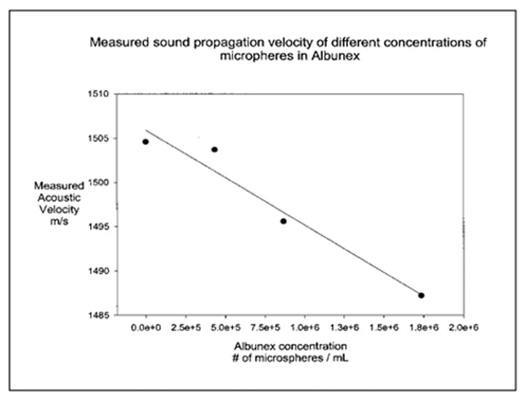
Sound velocity in an Albunex suspension as a function of concentration. The solid circles and the line represent the data points and the least squares regressions line, respectively.
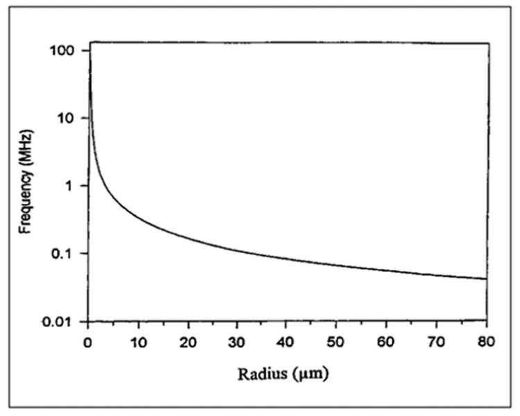
Computed resonance frequency versus the radius of a bubble.
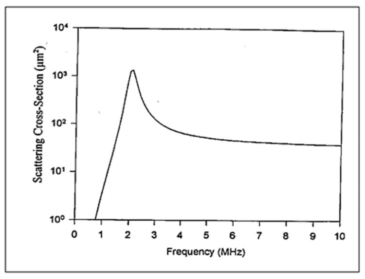
Computed scattering cross section of a bubble of 1.7 μm radius.
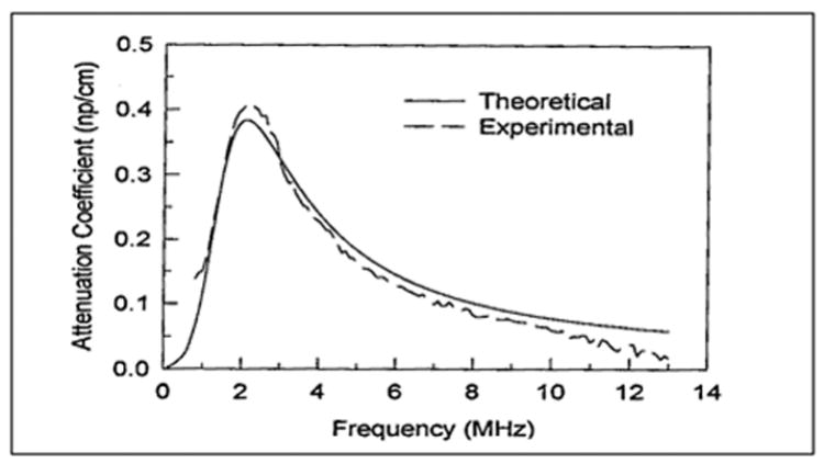
Experimental and theoretical results of attenuation coefficient of an Albunex suspension as a function of frequency. Mean diameter of Albunex = 3.15 μm.
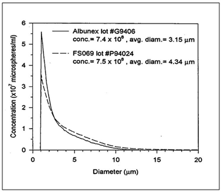
Size distributions of Albunex and FS069.
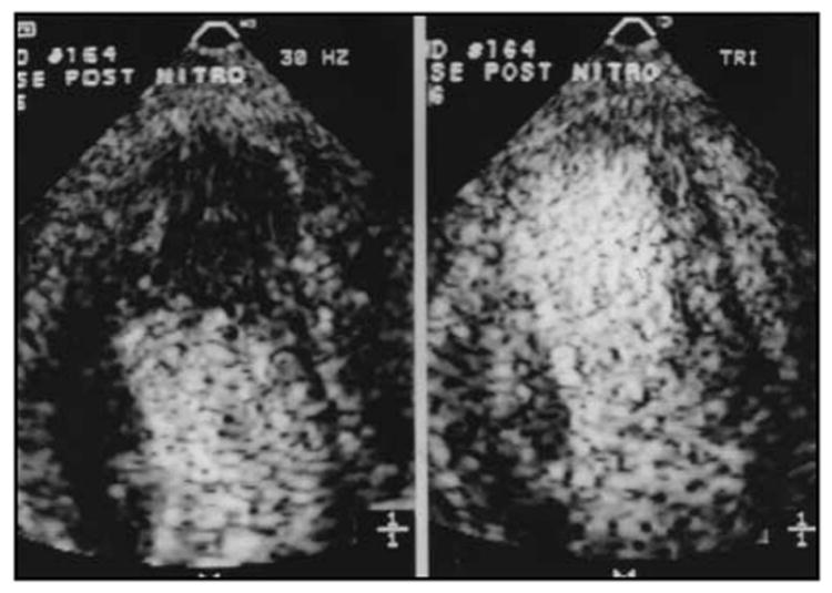
An example of the increase in myocardial contrast observed during continuous infusion of PESDA microbubbles when using a low frame rate (right panel) as opposed to conventional frame rates (left panel); the latter are usually > 30 Hz.
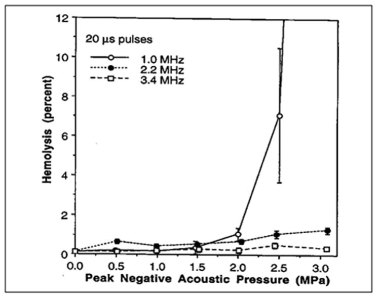
Hemolysis of whole human blood containing 3.6 V % Albunex and exposed with sample rotation to 20 μs pulses of 1.0, 2.2, or 3.4 MHz ultrasound. The duty factor was 0.01 and the total exposure duration 60 s. (Data from and reprinted by permission of Elsevier Science, from Brayman AA, et al: Hemolysis of 40% hematocrit, Albunex-supplemented human erythrocyte suspensions by intense pulsed ultrasound: Frequency, duty factor, pulse length and sample rotation dependence. Ultrasound Med Biol 23:1237, 1997b.)
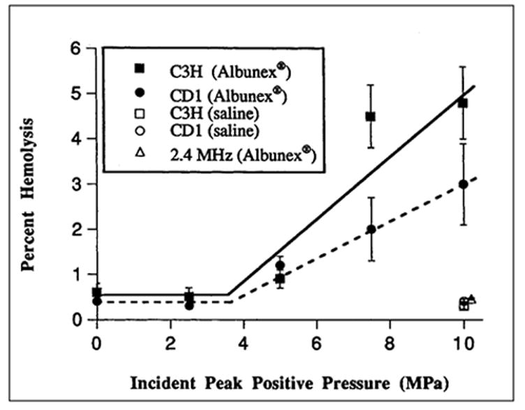
Hemolysis in vivo in mice exposed to pulsed ultrasound. Percent hemolysis is plotted as a function of peak positive pressure at the surface of the animal. Solid squares and circles are data for C3H and CD1 strains of mice, respectively, exposed to 1.2 MHz ultrasound and injected with Albunex. Open squares and circles are data for C3H and CD1 mice, respectively, exposed to 1.2 MHz ultrasound and injected with saline. Open triangle is datum for CD1 mice exposed to 2.4 MHz ultrasound and injected with Albunex. (Figure adapted from and reprinted by permission of Elsevier Science, from Dalecki D, et al: Remnants of Albunex nucleate acoustic cavitation. Ultrasound Med Biol 23:1405, 1997e). Data are presented as the mean percentage of hemolysis; error bars represent the standard error of the mean.
Similar articles
-
Section 5--nonthermal bioeffects in the absence of well-defined gas bodies.J Ultrasound Med. 2000 Feb;19(2):109-19, 154-68. doi: 10.7863/jum.2000.19.2.109. J Ultrasound Med. 2000. PMID: 10680617 Review.
-
Section 3--selected biological properties of tissues: potential determinants of susceptibility to ultrasound-induced bioeffects. American Institute of Ultrasound in Medicine.J Ultrasound Med. 2000 Feb;19(2):85-96, 154-68. doi: 10.7863/jum.2000.19.2.85. J Ultrasound Med. 2000. PMID: 10680615 Free PMC article. Review.
-
Section 4--bioeffects in tissues with gas bodies. American Institute of Ultrasound in Medicine.J Ultrasound Med. 2000 Feb;19(2):97-108, 154-68. doi: 10.7863/jum.2000.19.2.97. J Ultrasound Med. 2000. PMID: 10680616 Free PMC article. Review.
-
Overview of experimental studies of biological effects of medical ultrasound caused by gas body activation and inertial cavitation.Prog Biophys Mol Biol. 2007 Jan-Apr;93(1-3):314-30. doi: 10.1016/j.pbiomolbio.2006.07.027. Epub 2006 Aug 22. Prog Biophys Mol Biol. 2007. PMID: 16989895 Review.
-
Mechanical bioeffects of ultrasound.Annu Rev Biomed Eng. 2004;6:229-48. doi: 10.1146/annurev.bioeng.6.040803.140126. Annu Rev Biomed Eng. 2004. PMID: 15255769 Review.
Cited by
-
In vivo study of enhanced chemotherapy combined with ultrasound image-guided focused ultrasound (USgFUS) treatment for pancreatic cancer in a xenograft mouse model.Eur Radiol. 2018 Sep;28(9):3710-3718. doi: 10.1007/s00330-018-5355-9. Epub 2018 Mar 29. Eur Radiol. 2018. PMID: 29600477
-
Contrast Ultrasound Imaging of the Aorta Does Not Affect Progression of Atherosclerosis or Cardiovascular Biomarkers in ApoE-/- Mice.J Ultrasound Med. 2015 Jun;34(6):1115-22. doi: 10.7863/ultra.34.6.1115. J Ultrasound Med. 2015. PMID: 26014332 Free PMC article.
-
Neuroinflammation associated with ultrasound-mediated permeabilization of the blood-brain barrier.Trends Neurosci. 2022 Jun;45(6):459-470. doi: 10.1016/j.tins.2022.03.003. Epub 2022 Apr 20. Trends Neurosci. 2022. PMID: 35461727 Free PMC article. Review.
-
Lesions of ultrasound-induced lung hemorrhage are not consistent with thermal injury.Ultrasound Med Biol. 2006 Nov;32(11):1763-70. doi: 10.1016/j.ultrasmedbio.2006.06.012. Ultrasound Med Biol. 2006. PMID: 17112962 Free PMC article.
-
Vascular lesions and s-thrombomodulin concentrations from auricular arteries of rabbits infused with microbubble contrast agent and exposed to pulsed ultrasound.Ultrasound Med Biol. 2006 Nov;32(11):1781-91. doi: 10.1016/j.ultrasmedbio.2005.11.013. Ultrasound Med Biol. 2006. PMID: 17112964 Free PMC article.
References
-
- Abramowicz JS. Ultrasound contrast media and their use in obstetrics and gynecology. Ultrasound Med Biol. 1997;23:1287. - PubMed
-
- Abramowicz JS, Phillips DB, Jessee LN, et al. Enhanced blood flow visualization in the perfused human placenta by Albunex®, an ultrasound contrast medium. Placenta. 1996;17:A.21. - PubMed
-
- Achiron R, Lipitz S, Sivan E, et al. Sonohysterography for ultrasonographic evaluation of tamoxifen-associated cystic thickened endometrium. J Ultrasound Med. 1995;14:685. - PubMed
-
- Allahbadia GN. Fallopian tubes and ultrasonography: The Sion experience. Fertil Steril. 1992;58:901. - PubMed
-
- AIUM American Insitute of Ultrasound in Medicine Bioeffects Committee. Bioeffects and Safety of Diagnostic Ultrasound. Laurel, MD: American Institute of Ultrasound in Medicine; 1993.
Publication types
MeSH terms
Substances
Grants and funding
LinkOut - more resources
Full Text Sources
Other Literature Sources
