

Effects of atovaquone and diospyrin-based drugs on the cellular ATP of Pneumocystis carinii f. sp. carinii
- PMID: 10681344
- PMCID: PMC89752
- DOI: 10.1128/AAC.44.3.713-719.2000
Effects of atovaquone and diospyrin-based drugs on the cellular ATP of Pneumocystis carinii f. sp. carinii
Abstract
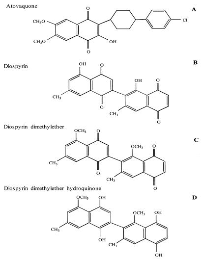
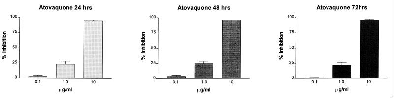
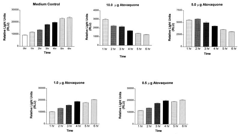
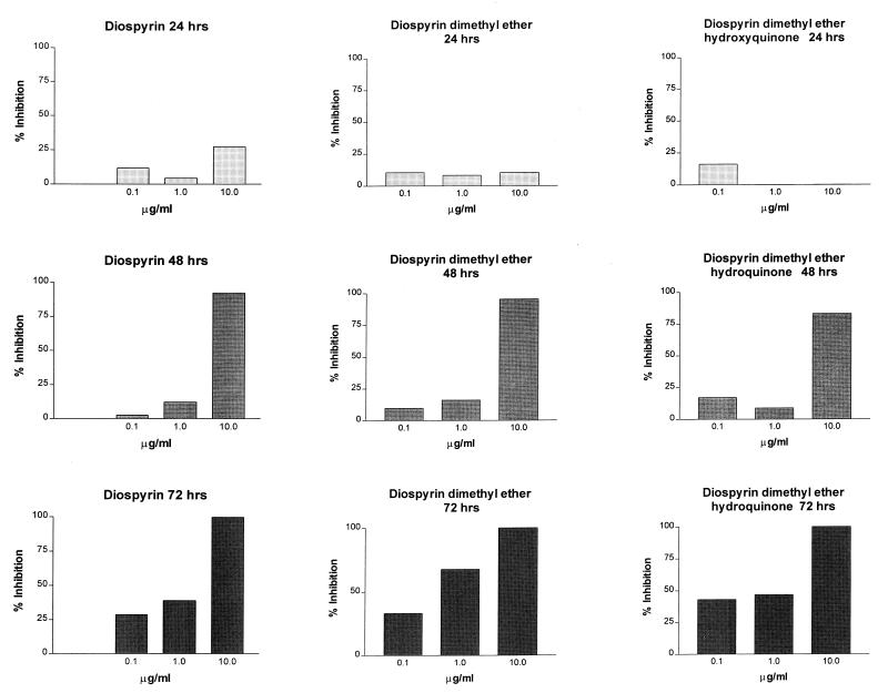
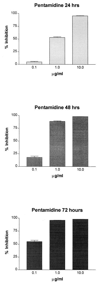
Atovaquone (also called Mepron, or 566C80) is a napthoquinone used for the treatment of infections caused by pathogens such as Plasmodium spp. and Pneumocystis carinii. The mechanism of action against the malarial parasite is the inhibition of dihydroorotate dehydrogenase (DHOD), a consequence of blocking electron transport by the drug. As an analog of ubiquinone (coenzyme Q [CoQ]), atovaquone irreversibly binds to the mitochondrial cytochrome bc(1) complex; thus, electrons are not able to pass from dehydrogenase enzymes via CoQ to cytochrome c. Since DHOD is a critical enzyme in pyrimidine biosynthesis, and because the parasite cannot scavenge host pyrimidines, the drug is lethal to the organism. Oxygen consumption in P. carinii is inhibited by the drug; thus, electron transport has also been identified as the drug target in P. carinii. However, unlike Plasmodium DHOD, P. carinii DHOD is inhibited only at high atovaquone concentrations, suggesting that the organism may salvage host pyrimidines and that atovaquone exerts its primary effects on ATP biosynthesis. In the present study, the effect of atovaquone on ATP levels in P. carinii was measured directly from 1 to 6 h and then after 24, 48, and 72 h of exposure. The average 50% inhibitory concentration after 24 to 72 h of exposure was 1.5 microgram/ml (4.2 microM). The kinetics of ATP depletion were in contrast to those of another family of naphthoquinone compounds, diospyrin and two of its derivatives. Whereas atovaquone reduced ATP levels within 1 h of exposure, the diospyrins required at least 48 h. After 72 h, the diospyrins were able to decrease ATP levels of P. carinii at nanomolar concentrations. These data indicate that although naphthoquinones inhibit the electron transport chain, the molecular targets in a given organism are likely to be distinct among members of this class of compounds.
Figures
Similar articles
-
Effects of atovaquone and diospyrin-based drugs on ubiquinone biosynthesis in Pneumocystis carinii organisms.Antimicrob Agents Chemother. 2000 Jan;44(1):14-8. doi: 10.1128/AAC.44.1.14-18.2000. Antimicrob Agents Chemother. 2000. PMID: 10602716 Free PMC article.
-
Effects of atovaquone and other inhibitors on Pneumocystis carinii dihydroorotate dehydrogenase.Antimicrob Agents Chemother. 1995 Feb;39(2):325-8. doi: 10.1128/AAC.39.2.325. Antimicrob Agents Chemother. 1995. PMID: 7726490 Free PMC article.
-
Are cytochrome b gene mutations the only cause of atovaquone resistance in Pneumocystis?Drug Resist Updat. 2001 Oct;4(5):322-9. doi: 10.1054/drup.2001.0221. Drug Resist Updat. 2001. PMID: 11991686 Review.
-
Ubiquinone synthesis in mitochondrial and microsomal subcellular fractions of Pneumocystis spp.: differential sensitivities to atovaquone.Eukaryot Cell. 2005 Aug;4(8):1483-92. doi: 10.1128/EC.4.8.1483-1492.2005. Eukaryot Cell. 2005. PMID: 16087753 Free PMC article.
-
Pneumocystis carinii pneumonia: the status of Pneumocystis biochemistry.Int J Parasitol. 1998 Jan;28(1):65-84. doi: 10.1016/s0020-7519(97)00179-3. Int J Parasitol. 1998. PMID: 9504336 Review.
Cited by
-
Biofilm formation by Pneumocystis spp.Eukaryot Cell. 2009 Feb;8(2):197-206. doi: 10.1128/EC.00202-08. Epub 2008 Sep 26. Eukaryot Cell. 2009. PMID: 18820078 Free PMC article.
-
Sterol metabolism in the opportunistic pathogen Pneumocystis: advances and new insights.Lipids. 2004 Aug;39(8):753-61. doi: 10.1007/s11745-004-1292-5. Lipids. 2004. PMID: 15638243 Review.
-
Susceptibility of Pneumocystis to echinocandins in suspension and biofilm cultures.Antimicrob Agents Chemother. 2011 Oct;55(10):4513-8. doi: 10.1128/AAC.00017-11. Epub 2011 Jul 25. Antimicrob Agents Chemother. 2011. PMID: 21788469 Free PMC article.
-
Axenic Long-Term Cultivation of Pneumocystis jirovecii.J Fungi (Basel). 2023 Sep 1;9(9):903. doi: 10.3390/jof9090903. J Fungi (Basel). 2023. PMID: 37755011 Free PMC article.
-
Gene arrays at Pneumocystis carinii telomeres.Genetics. 2005 Aug;170(4):1589-600. doi: 10.1534/genetics.105.040733. Epub 2005 Jun 18. Genetics. 2005. PMID: 15965256 Free PMC article.
References
-
- Anabwani G, Canfield C J, Hutchinson D B. Combination atovaquone and proguanil hydrochloride vs. halofantrine for treatment of acute Plasmodium falciparum malaria in children. Pediatr Infect Dis J. 1999;18:456–461. - PubMed
-
- Cirioni O, Giacometti A, Scalise G. In vitro activity of atovaquone, sulphamethoxazole and dapsone alone and combined with inhibitors of dihydrofolate reductase and macrolides against Pneumocystis carinii. J Antimicrob Chemother. 1997;39:45–51. - PubMed
-
- Comley J C W, Mullin R J, Wolfe L A, Hanlon M H, Ferone R. A radiometric method for objectively screening large numbers of compounds against Pneumocystis carinii in vitro. J Protozool. 1991;38:144S–146S. - PubMed
Publication types
MeSH terms
Substances
Grants and funding
LinkOut - more resources
Full Text Sources
Other Literature Sources
Research Materials
Miscellaneous
