. 2000 Feb 29;97(5):2235-40.
doi: 10.1073/pnas.040549997.

Crystal structure of the zymogen form of the group A Streptococcus virulence factor SpeB: an integrin-binding cysteine protease

Affiliations

Crystal structure of the zymogen form of the group A Streptococcus virulence factor SpeB: an integrin-binding cysteine protease

T F Kagawa et al. Proc Natl Acad Sci U S A. .

Abstract

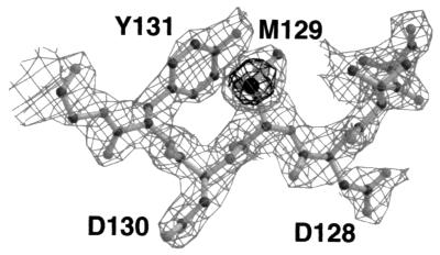
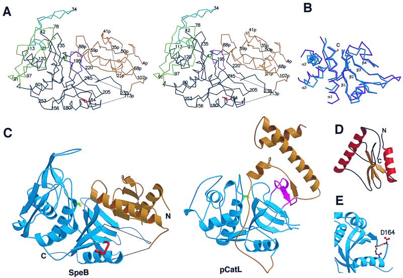
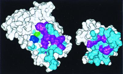
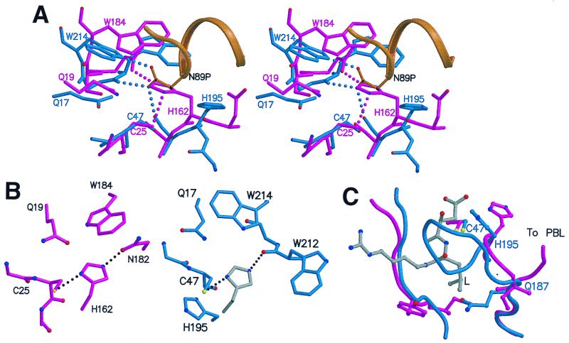
Pathogenic bacteria secrete protein toxins that weaken or disable their host, and thereby act as virulence factors. We have determined the crystal structure of streptococcal pyrogenic exotoxin B (SpeB), a cysteine protease that is a major virulence factor of the human pathogen Streptococcus pyogenes and participates in invasive disease episodes, including necrotizing fasciitis. The structure, determined for the 40-kDa precursor form of SpeB at 1.6-A resolution, reveals that the protein is a distant homologue of the papain superfamily that includes the mammalian cathepsins B, K, L, and S. Despite negligible sequence identity, the protease portion has the canonical papain fold, albeit with major loop insertions and deletions. The catalytic site differs from most other cysteine proteases in that it lacks the Asn residue of the Cys-His-Asn triad. The prosegment has a unique fold and inactivation mechanism that involves displacement of the catalytically essential His residue by a loop inserted into the active site. The structure also reveals the surface location of an integrin-binding Arg-Gly-Asp (RGD) motif that is a feature unique to SpeB among cysteine proteases and is linked to the pathogenesis of the most invasive strains of S. pyogenes.


Figures

Figure 1
Figure 1
Section of the 2.4-Å experimentally phased, NCS-averaged electron density map, contoured at 1.5 σ and 6 σ.
Figure 2
Figure 2
Structure of SpeB and comparison with other papain-family proteins. (A) Stereo Cα plot of SpeB. The prosegment is colored gold and the protease domains black, except for the two N-domain insertions 19–42 (the finger loop, blue) and 66–113 (green) and the truncated β1-β2 C-domain loop 187–195 (magenta). Asp-164 of the RGD loop is red and the Ser-47 side chain yellow. Dotted lines indicate flexible regions. (B) Superposition of the core secondary structures of mSpeB (blue) and actinidin (magenta). The location of the catalytic cysteine (C) is shown. (C) Ribbon diagrams of SpeB (Left) and procathepsin L (Right), showing their similar protease domains (blue) and different prosegments (gold). The RGD loop of SpeB is red. The PBL, present in other papain-family enzymes but not SpeB, is colored magenta in procathepsin L. (D) Folding of the prosegment. (E). RGD loop (gold) of SpeB, showing the exposed Asp-164. These and other figures were produced with molscript (33) and raster3d (34).
Figure 3
Figure 3
Association of the SpeB prosegment and protease domains. The predominantly hydrophobic contact surfaces of the prosegment (Right) and protease (Left) are colored blue, with aromatic residues in purple. His-195 and Ser(Cys)-47 are blue and green, respectively. Hydrophobic residues involved include Phe-61p, Pro-71p, Tyr-76p, Phe-82p, Ile-90p, Phe-93p, Met-94p, and Tyr-97p from the prosegment and Tyr-186, Phe-197, Phe-207, Trp-212, Trp-214, Val-217, Gly-220, Phe-221, Phe-222, and Ala-226 from the protease.
Figure 4
Figure 4
Active site region of SpeB, compared with actinidin. (A) Stereo view of zSpeB (blue and gold) superimposed on actinidin (magenta). In zSpeB, the insertion of Asn-89p from the prosegment (gold) displaces His-195 from the catalytically competent position of His-162 in actinidin. Hydrogen bonds made by Asn-89p in SpeB and His-162 in actinidin are shown by dotted lines. (B) The Cys-His-Asn triad in actinidin (Left) compared with the putative Cys-His-(O=C) triad in mSpeB (Right). For mSpeB we assume that His-195 rotates from its position in the zymogen to a putative catalytic position shown in gray. (C) The S2 binding pocket. The leucyl side chain (L) of an inhibitor E64 (gray) is shown as it binds in actinidin (43). In zSpeB (blue) the loop 187–195 blocks the pocket; in actinidin (magenta) the chain goes in a different direction to form the PBL and the pocket is open.

References

    1. Quinn R W. Rev Infect Dis. 1989;11:928–953. - PubMed
    1. Proft T, Moffatt S L, Berkhan C J, Fraser J D. J Exp Med. 1999;189:89–101. - PMC - PubMed
    1. Musser J M. In: Superantigens: Molecular Biology, Immunology, and Relevance to Human Disease. Leung D Y M, Huber B T, Schlievert P M, editors. New York: Dekker; 1997. pp. 281–310.
    1. Musser J M, Hauser A R, Kim M, Schlievert P M, Nelson K, Selander R K. Proc Natl Acad Sci USA. 1991;88:2668–2672. - PMC - PubMed
    1. Kapur V, Topouzis S, Majesky M W, Li L-L, Hamrick M R, Hamill R J, Patti J M, Musser J M. Microb Pathog. 1993;15:327–346. - PubMed

Publication types

MeSH terms

Associated data

LinkOut - more resources