. 2000 Feb 29;97(5):2157-62.
doi: 10.1073/pnas.040568397.

Toward Anopheles transformation: Minos element activity in anopheline cells and embryos

Affiliations

Toward Anopheles transformation: Minos element activity in anopheline cells and embryos

F Catteruccia et al. Proc Natl Acad Sci U S A. .

Erratum in

  • Proc Natl Acad Sci U S A 2000 May 23;97(11):6236

Abstract

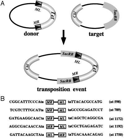
The ability of the Minos transposable element to function as a transformation vector in anopheline mosquitoes was assessed. Two recently established Anopheles gambiae cell lines were stably transformed by using marked Minos transposons in the presence of a helper plasmid expressing transposase. The markers were either the green fluorescent protein or the hygromycin B phosphotransferase gene driven by the Drosophila Hsp70 promoter. Cloning and sequencing of the integration sites demonstrated that insertions in the cell genome occurred through the action of Minos transposase. Furthermore, an interplasmid transposition assay established that Minos transposase is active in the cytoplasmic environment of Anopheles stephensi embryos: interplasmid transposition events isolated from injected preblastoderm embryos were identified as Minos transposase-mediated integrations, and no events were recorded in the absence of an active transposase. These results demonstrate that Minos vectors are suitable candidates for germ-line transformation of anopheline mosquitoes.


Figures

Figure 1
Figure 1
Diagram of the excision assay and results from three A. gambiae cell lines (4a-2, Sua 4.0, and Sua 5.1*) as well as the mbn2 hemocyte cell line of D. melanogaster (25), transformed with donor plasmid with (+) or without (−) a transposase-producing plasmid. Excision results in a DNA circle from sequences outside the Minos ends. Primers annealing to sequences within the circle (black arrows) can drive a PCR, resulting in a 167-bp excision band in + but not in − lanes. Lane C shows a control PCR without template. The doublet of bands at ca. 400 bp is derived from ectopic priming from donor plasmid sequences and is competed by the specific priming (compare lanes + and −).
Figure 2
Figure 2
(A) Schematic representation of plasmid pMinHyg used for genetic transformation of Sua 4.0 cells. The hatched boxes represent Minos sequences located internally to the Minos ends (boxed arrowheads). Minos sequences were interrupted by the insertion of an Hsp70-hygromycin B phosphotransferase fusion gene (Hsp70-Hyg), and a stuffer DNA sequence that includes a 2.6-kb fragment of the D. melanogaster act5C promoter and a 0.8-kb fragment of the Hsp70 terminator (marked as A–G). The ampicillin resistance gene derived from plasmid pTZ18R also is indicated. The black bars delineate the probes used for Southern analysis. N, NotI; E, EcoRI; H, HincII. (B) Southern analysis of HincII-digested genomic DNA of the Sua 4.0 clones hybridized with probe M. B1, C1, C2, B11, B12, C12, F1, and D2 represent eight different hygromycin-resistant clones of Sua 4.0 cells, transfected with a mixture of the pMiHyg plasmid and the helper plasmid pHSS6hsMi20. The probe verifies the presence of Minos transposon sequences in the genomic DNA of these stable tranfected cells. Clone B12 is a spontaneously resistant clone that lacks insertions and is included as a negative control. The arrowheads indicate bands of size and intensity consistent with expectation for illegitimate integrations of pMiHyg plasmid concatamers in the chromosomes of the cells. * indicates a band expected for concatamers of the helper plasmid. (C) Southern analysis of the same clones digested with HincII restriction enzyme and probed with probe Amp. A 2.9-kb band indicates the presence of sequences derived from the plasmid backbone in all of the clones except F1.
Figure 3
Figure 3
(Aa) Schematic representation of the plasmid pLHGR used to transfect the Sua 5.1* cells. Between the Minos ends (boxed arrowheads), an Hsp70-GFP fusion gene and a whole Bluescript plasmid (PS) were incorporated. Outside the Minos ends, D. hydei genomic sequences and a DNA fragment corresponding to the tetracycline resistance gene generate a stuffer between the two ends. (Ab) Predicted legitimate insertion of the above transposon in genomic DNA (crosshatched lines). P, PstI; S, SacII restriction sites. Letters in parenthesis indicate the same restriction sites that may be present in the A. gambiae flanking genomic DNA. (Ac) Predicted structure of rescued plasmids corresponding to canonical Minos insertions in the genome of Sua 5.1* cells. Such plasmids should contain a common Bluescript-bearing DNA fragment (PS) linked to a variable size fragment extending into A. gambiae flanking genomic DNA. (B) Comparison of histograms of cell populations derived from Sua 5.1* cells initially transfected with plasmid pLHGR in the presence (+H) or the absence (−H) of helper plasmid. The transfected cells were expanded and resorted after 40 days posttransfection. A fluorescent cell fraction could be sorted from the +H cells. The horizontal axis corresponds to increasing fluorescence intensity expressed on a logarithmic scale. The vertical axis corresponds to cell counts per fluorescence channel. (Inset) A pair of fluorescent cells. (C) Restriction patterns of rescued plasmids representing 10 different Minos-mediated insertion events. The plasmids were digested with a PstI/SacII enzyme combination. A common Bluescript restriction band, 2.9 kb long (PS), is present in all of the plasmids, together with bands of variable size deriving from the flanking genomic DNA. A second variable band in the same lane indicates the presence of a SacII site in the flanking DNA (see Ac). In the case of E6, the second band was caused by a PstI fragment derived from the X chromosome that was trapped in the plasmid during the rescue procedure.
Figure 4
Figure 4
(A) Sequences of the Minos insertion sites in the genome of Sua 5.1* and Sua 4.0 cells. Chromosomal flanking sequences are represented with capital letters in italics. Small lettering represents the sequences of the Minos end. The expected TA dinucleotide of the insertion site is shown in bold. The chromosomal divisions and subdivisions from which the flanking sequences were derived are indicated with the chromosomal arm listed in parenthesis. (B) Typical results of determining the location of origin of the rescued genomic fragments by in situ localization to polytene chromosomes of the Suakoko mosquito strain.
Figure 5
Figure 5
(A) Schematic representation of interplasmid transposition events. Transposition of a Minos transposon from the donor plasmid into the sucrase gene carried by the target plasmid results in a combined plasmid that can be selected by using a triple selection scheme (Chl, chloramphenicol; Tet, tetracycline; SacRB, sucrase). (B) Sequences flanking the ends of Minos insertions in the sucrase gene, in events that occurred in A. stephensi embryos. The nucleotide positions in the sucrase gene (Genbank accession nos. XO2730 and KO1987), at which the insertion took place are indicated. The orientation of the insertion relative to the sucrase sequence also is indicated (ML-MR or MR-ML).

References

    1. Miller L H, Sakai R K, Romans P, Gwadz R W, Kantoff P, Coon H G. Science. 1987;237:779–781. - PubMed
    1. Loukeris T G, Livadaras I, Arca B, Zabalou S, Savakis C. Science. 1995;270:2002–2005. - PubMed
    1. Coates C J, Jasinskiene N, Miyashiro L, James A A. Proc Natl Acad Sci USA. 1998;95:3748–3751. - PMC - PubMed
    1. Jasinskiene N, Coates C J, Benedict M Q, Cornel A J, Rafferty C S, James A A, Collins F H. Proc Natl Acad Sci USA. 1998;95:3743–3747. - PMC - PubMed
    1. Lidholm D A, Lohe A R, Hartl D L. Genetics. 1993;134:859–868. - PMC - PubMed

Publication types