. 2000 Feb 29;97(5):2081-6.
doi: 10.1073/pnas.050316997.

Low dielectric response in enzyme active site

Affiliations

Low dielectric response in enzyme active site

E L Mertz et al. Proc Natl Acad Sci U S A. .

Abstract

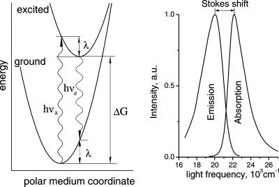
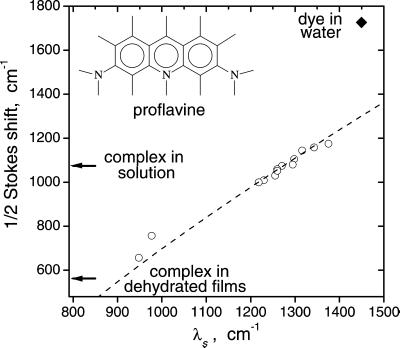
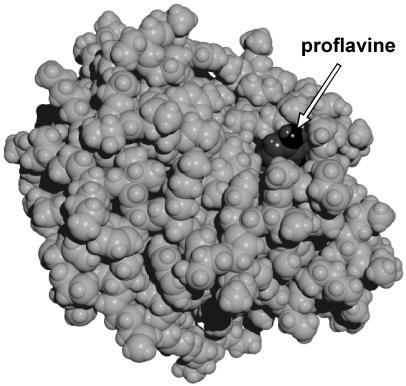
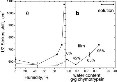
The kinetics of charge transfer depend crucially on the dielectric reorganization of the medium. In enzymatic reactions that involve charge transfer, atomic dielectric response of the active site and of its surroundings determines the efficiency of the protein as a catalyst. We report direct spectroscopic measurements of the reorganization energy associated with the dielectric response in the active site of alpha-chymotrypsin. A chromophoric inhibitor of the enzyme is used as a spectroscopic probe. We find that water strongly affects the dielectric reorganization in the active site of the enzyme in solution. The reorganization energy of the protein matrix in the vicinity of the active site is similar to that of low-polarity solvents. Surprisingly, water exhibits an anomalously high dielectric response that cannot be described in terms of the dielectric continuum theory. As a result, sequestering the active site from the aqueous environment inside low-dielectric enzyme body dramatically reduces the dielectric reorganization. This reduction is particularly important for controlling the rate of enzymatic reactions.


Figures

Figure 1
Figure 1
A schematic representation of the energetics of electronic transitions in a dye in a polarizable medium (Left) and the corresponding absorption and emission spectra for proflavine dye in a polar medium (Right). The x axis on the left is a generalized classical coordinate describing, e.g., the orientation of surrounding dipoles in the medium. The light absorption (hνa) and emission (hνe) transitions are shown at the most probable coordinates that are expected to determine the maxima of the corresponding spectra.
Figure 2
Figure 2
Semiquantitative evaluation of medium reorganization energy, λs, from the measured Stokes shift of proflavine: ○, calibration data for polar aprotic solvents with known dielectric properties (24) vs. calculated, scaled λs; ♦, the Stokes shift in water vs. λs calculated at ɛs = 80 and ɛo = 1.77. The dashed line shows the fit to Eq. 2 of the data for aprotic solvents (24). This fit can be used as a calibration curve to estimate λs at the active site of chymotrypsin from the measured Stokes shifts of the dye–enzyme complex (shown by arrows). The lowest and uppermost open circles are, respectively, for dichloromethane and acetonitrile. Quantitative evaluation of λs with correction for the spectral moments is described in the text.
Figure 3
Figure 3
The proflavine–chymotrypsin complex. The arrow points to the edge of proflavine molecule. The structure was obtained by 10-ps molecular dynamics simulations. Calculations were performed within hyper chem 5.0 (Hypercube, Gainesville, FL) package using the amber (64) force field with 4,000 water molecules in the periodic box of 56 × 50 × 56 Å with the time step of 1 fs. Partial charges of proflavine were obtained from CNDO/2 semiempirical quantum chemical calculations. Other force field parameters were taken from amber parameters corresponding to similar atoms and chemical bonds of nucleotide bases. X-ray coordinates of chymotrypsin atoms with added hydrogen atoms and equilibrated for 20-ps molecular dynamics run without the dye were taken as the initial configuration of the protein.
Figure 4
Figure 4
(a) Stokes shift of proflavine in chymotrypsin films vs. humidity. Lower curve (□) is for films infused with chloroform. (b) Stokes shift in films without chloroform vs. water content. The water content was calculated from humidity by using the water adsorption isotherm on chymotrypsin measured in refs. and .
Figure 5
Figure 5
Circular dichroism spectra of chymotrypsin in solution and in protein films at fixed humidity.

References

    1. Marcus R A, Sutin N. Biochim Biophys Acta. 1985;811:265–322.
    1. Krishtalik L I. Mol Biol. 1974;8:91–99. - PubMed
    1. Krishtalik L I. Mol Biol. 1979;13:577–581. - PubMed
    1. Krishtalik L I. J Theor Biol. 1980;86:757–771. - PubMed
    1. Bailey S. Trans Faraday Soc. 1951;47:509–517.

Publication types