. 2000 Feb 29;97(5):2017-22.
doi: 10.1073/pnas.050417797.

Electrostatic stress in catalysis: structure and mechanism of the enzyme orotidine monophosphate decarboxylase

Affiliations

Electrostatic stress in catalysis: structure and mechanism of the enzyme orotidine monophosphate decarboxylase

N Wu et al. Proc Natl Acad Sci U S A. .

Abstract

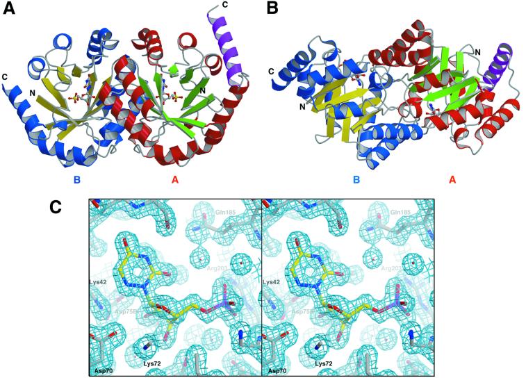
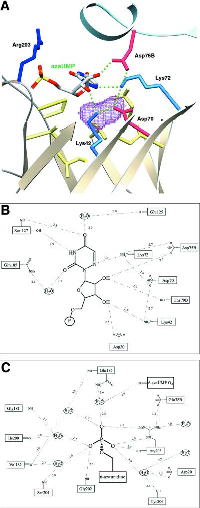
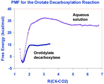
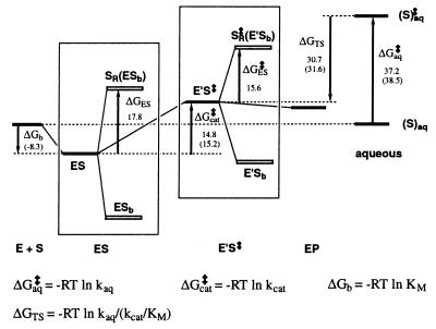
Orotidine 5'-monophosphate decarboxylase catalyzes the conversion of orotidine 5'-monophosphate to uridine 5'-monophosphate, the last step in biosynthesis of pyrimidine nucleotides. As part of a Structural Genomics Initiative, the crystal structures of the ligand-free and the6-azauridine 5'-monophosphate-complexed forms have been determined at 1.8 and 1.5 A, respectively. The protein assumes a TIM-barrel fold with one side of the barrel closed off and the other side binding the inhibitor. A unique array of alternating charges (Lys-Asp-Lys-Asp) in the active site prompted us to apply quantum mechanical and molecular dynamics calculations to analyze the relative contributions of ground state destabilization and transition state stabilization to catalysis. The remarkable catalytic power of orotidine 5'-monophosphate decarboxylase is almost exclusively achieved via destabilization of the reactive part of the substrate, which is compensated for by strong binding of the phosphate and ribose groups. The computational results are consistent with a catalytic mechanism that is characterized by Jencks's Circe effect.


Figures

Figure 1
Figure 1
Ribbon diagram of the ODCase dimer. (A) The 2-fold axis runs vertically. Monomer A is colored in red and green (for helices and strands of β-sheets, respectively) while monomer B is colored in blue and yellow. The extra helix from the pET15b vector is colored in purple. The barrel opening is facing up in monomer B. (B) Side view of the dimer. The 2-fold axis is now perpendicular to the page. (C) Stereoview of the active site with 2Fo-Fc map (contoured at 1.5 σ). The map was calculated before 6-azaUMP was added to the model, providing a totally unbiased representation of the bound inhibitor.
Figure 2
Figure 2
(A) Structural view of the active site, with the important elements and interactions of catalysis. Monomer A is colored in light gold and monomer B in pale blue. Even with 6-azaUMP bound, there is a cavity in the active site [purple contour, calculated with a van der Waals probe of 1.2 Å radius. This cavity is big enough to accommodate the C6 carboxylate of OMP or the product CO2. Hydrophobic residues lining the active site are shown in pale yellow. (B) Schematic view of the interactions between the active site and 6-azauridine. (C) Schematic view of ionic interactions and H-bonds associated with the phosphate group. All of the distances given are averages of values found in the four molecules in the asymmetric unit. The various corresponding distances differ by less than 0.3 Å from each other.
Figure 3
Figure 3
Computed free energy reaction profiles for the decarboxylation reactions of 1-methylorotate to 1-methyluracil anion and CO2 in water, and OMP to form the UMP anion and CO2 in ODCase. PMF, potential of mean force; R, the distance between the C6-atom of orotidine and the carbon of the leaving group, CO2.
Figure 4
Figure 4
A schematic representation of the Circe-effect energy-decomposition in the GS and TS. Computed binding and activation free energies (kcal/mol) are shown, along with the corresponding experimental values given in parentheses. The hidden thermodynamics of the overall observed binding energies for GS is revealed by considering the reactive orotate group (SR) and the ribose and phosphate binding part (Sb) of the substrate separately. A similar energy decomposition is also shown for the TS. KM is the Michelis-Menten dissociation constant, and kcat and kaq are rate constants for the catalyzed and noncatalyzed aqueous reaction, respectively. KTS = kaq/(kcat/KM) is an apparent equilibrium constant for the dissociation of the transition state from the enzyme (34). E and E′ denote different conformational states of the enzyme when the substrate is at the GS and TS, respectively.

References

    1. Lieberman I, Kornberg A, Simms E S. J Biol Chem. 1955;215:403–415. - PubMed
    1. Radzicka A, Wolfenden R. Science. 1995;267:90–93. - PubMed
    1. Beak P, Siegel B. J Am Chem Soc. 1976;98:3601–3606. - PubMed
    1. Levine H L, Brody R S, Westheimer F H. Biochemistry. 1980;19:4993–4999. - PubMed
    1. Acheson S A, Bell J B, Jones M E, Wolfenden R. Biochemistry. 1990;29:3198–3202. - PubMed

Publication types

Substances