. 2000 Feb 29;97(5):2087-92.
doi: 10.1073/pnas.030539197.

Compartmentation protects trypanosomes from the dangerous design of glycolysis

Affiliations

Compartmentation protects trypanosomes from the dangerous design of glycolysis

B M Bakker et al. Proc Natl Acad Sci U S A. .

Abstract

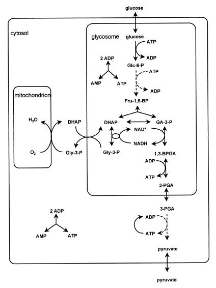
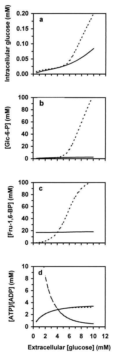
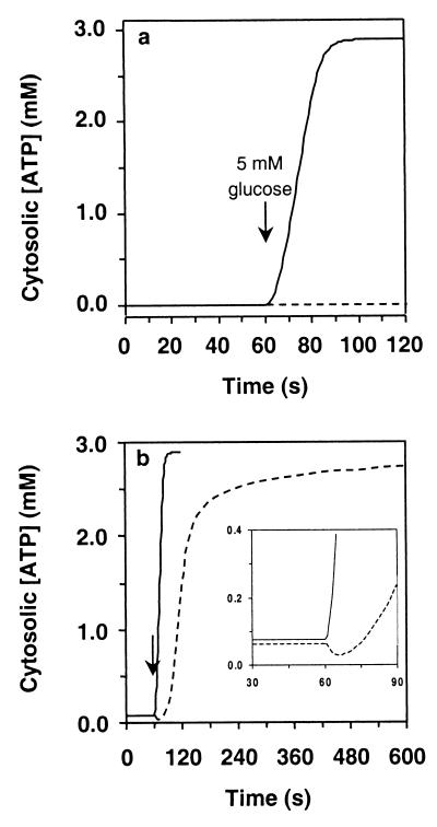
Unlike in other organisms, in trypanosomes and other Kinetoplastida the larger part of glycolysis takes place in a specialized organelle, called the glycosome. At present it is impossible to remove the glycosome without changing much of the rest of the cell. It would seem impossible, therefore, to assess the metabolic consequences of this compartmentation. Therefore, we here develop a computer experimentation approach, which we call computational cell biology. A validated molecular kinetic computer replica was built of glycolysis in the parasite Trypanosoma brucei. Removing the glycosome membrane in that replica had little effect on the steady-state flux, which argues against the prevalent speculation that glycosomes serve to increase flux by concentrating the enzymes. Removal of the membrane did cause (i) the sugar phosphates to rise to unphysiologically high levels, which must have pathological effects, and (ii) a failure to recover from glucose deprivation. We explain these effects on the basis of the biochemical organization of the glycosome. We conclude (i) that the glycosome protects trypanosomes from the negative side effects of the "turbo" structure of glycolysis and (ii) that computer experimentation based on solid molecular data is a powerful tool to address questions that are not, or not yet, accessible to experimentation.


Figures

Figure 1
Figure 1
A scheme of glycolysis in bloodstream-form Trypanosoma brucei. Solid lines represent reactions catalyzed by a single enzyme; dashed lines represent multiple sequential reactions. Glc-6-P, glucose 6-phosphate; Fru-1,6-BP, fructose 1,6-bisphosphate; GA-3-P, glyceraldehyde 3-phosphate; 1,3-BPGA, 1,3-bisphosphoglycerate; 3-PGA, 3-phosphoglycerate; DHAP, dihydroxyacetone phosphate; Gly-3-P, glycerol 3-phosphate.
Figure 2
Figure 2
In trypanosomes lacking the glycosomal membrane, hexose phosphates accumulate to extremely high levels. The steady-state intracellular concentrations of glucose (a), Glc-6-P (b), and Fru-1,6-BP (c) were calculated both for the presence (solid lines) and for the absence (dashed lines) of the glycosomal membrane. (d) Glycosomal (long-dashed line) and (solid line) cytosolic [ATP]/[ADP] ratio for if the glycosome is present and the cytosolic [ATP]/[ADP] ratio for if there is no glycosome (short-dashed line).
Figure 3
Figure 3
The glycosome helps trypanosomes to recover from a low blood-glucose level when returning to a normal one. Trypanosomes consuming glucose in a steady state at 0.01 mM (a) or 0.05 mM (b) extracellular glucose, were given 5 mM glucose after 60 s. The cytosolic ATP concentration was monitored, both in the model with the glycosome (solid line) and in the model without the glycosome (dashed line). (Inset) Part from 30 to 90 s enlarged.

References

    1. Opperdoes F R, Borst P. FEBS Lett. 1977;80:360–364. - PubMed
    1. Tetley L, Vickerman K. J Microsc. 1991;162:83–90. - PubMed
    1. Opperdoes F R, Baudhuin P, Coppens I, De Roe C, Edwards S W, Weijers P J, Misset O. J Cell Biol. 1984;98:1178–1184. - PMC - PubMed
    1. Visser N, Opperdoes F R, Borst P. Eur J Biochem. 1981;118:521–526. - PubMed
    1. Opperdoes F R. Annu Rev Microbiol. 1987;41:127–151. - PubMed

Publication types

MeSH terms

LinkOut - more resources