. 2000 Feb 29;97(5):2391-6.
doi: 10.1073/pnas.040565697.

The cellulose synthase gene of Dictyostelium

Affiliations

The cellulose synthase gene of Dictyostelium

R L Blanton et al. Proc Natl Acad Sci U S A. .

Abstract

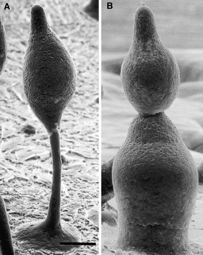
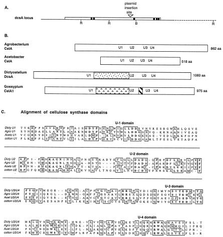
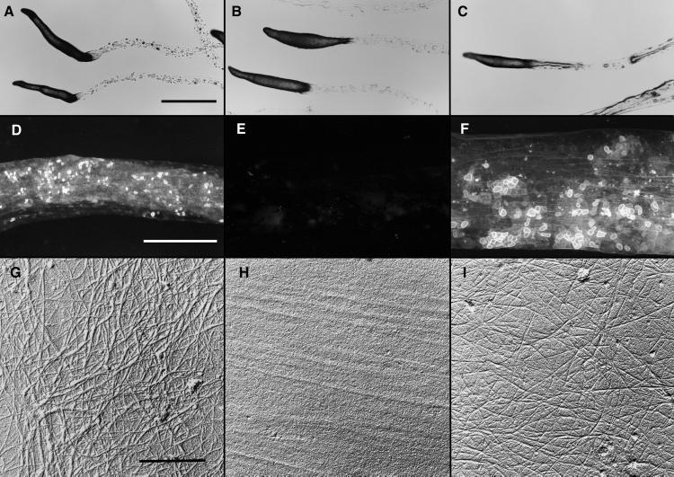
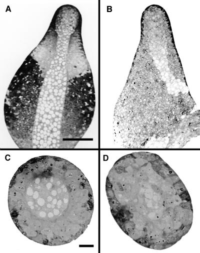
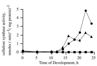
Cellulose is a major component of the extracellular matrices formed during development of the social amoeba, Dictyostelium discoideum. We isolated insertional mutants that failed to accumulate cellulose and had no cellulose synthase activity at any stage of development. Development proceeded normally in the null mutants up to the beginning of stalk formation, at which point the culminating structures collapsed onto themselves, then proceeded to attempt culmination again. No spores or stalk cells were ever made in the mutants, with all cells eventually lysing. The predicted product of the disrupted gene (dcsA) showed significant similarity to the catalytic subunit of cellulose synthases found in bacteria. Enzyme activity and normal development were recovered in strains transformed with a construct expressing the intact dcsA gene. Growing amoebae carrying the construct accumulated the protein product of dcsA, but did not make cellulose until they had developed for at least 10 hr. These studies show directly that the product of dcsA is necessary, but not sufficient, for synthesis of cellulose.


Figures

Figure 1
Figure 1
Scanning electron micrographs of developing wild-type and cellulose-deficient strains. (A) Developing fruiting body of the wild type after 18 hr of development. Note the well-defined stalk and basal disk. (B) Developing fruiting body of the cellulose-deficient strain DG1099 after 18 hr of development. Note the absence of a stalk or basal disk. The apical-most mass is similar in appearance to the cell mass in A. The multiple lobes seen here are characteristic of the mutant strains, giving them the appearance of snowmen. (Bar = 50 μm.)
Figure 2
Figure 2
Structure of the dcsA gene and comparison of its product, DcsA, with related proteins. (A) The region flanking DpnII restriction site (▿), where plasmids were found to have inserted in strains DG1099 and DG1128, contains a single, large ORF of 3.2 kb (open box). Pertinent restriction sites (RI, EcoRI; B, BglII) are marked. Sequences encoding hydrophobic domains of ≥20 aa are shown as solid boxes. (B) The predicted product, DcsA, is most similar to the cellulose synthases of A. tumefaciens and A. xylinum and could be aligned on the basis of conserved motifs U2–U4, which are marked at their position within each protein. An insertion occurred between the U1 and U2 motifs in both DcsA and the putative cellulose synthase of cotton, G. hirsutum, CelA1. However, the sequence of the insert is not conserved between these organisms. (C) Detailed comparison of the amino acid sequences surrounding the conserved motifs shows extended similarities especially between the established bacterial cellulose synthases and DcsA.
Figure 3
Figure 3
Expression of dcsA. (A) RNA was prepared at 4-hr intervals during early development and 2-hr intervals throughout late development of wild type (AX4), the dcsA strain (DG1128), and the rescued strain (TL128). (B) An antibody was prepared against a peptide corresponding to the U2 region of DcsA. Total cellular proteins were prepared at 4-hr intervals during early development and 2-hr intervals throughout late development of wild type (AX4), the dcsA strain (DG1128), and the rescued strain (TL128).
Figure 4
Figure 4
Slime trails produced by slugs of wild-type, dcsA, and the dcsA rescued strains. Migrating slugs were phototactically directed to migrate across 2% water agar dishes. They were photographed in place (AC) or the trails were collected onto coverslips (DF) or electron microscope specimen grids (GI). (A–C) All strains formed normal-appearing slugs that migrated phototactically and left behind slime trails. (Bar = 1 mm.) (D–F) The trails left behind wild-type slugs (D), and those of the rescued strain TL128 (F) fluoresced brightly whereas there was only background fluorescence from the trails left behind the dcsA strain DG1128 (E). (Bar = 100 nm.) (G–I) Slime trails collected on electron microscope specimen grids were treated with Proteinase K (to remove obscuring proteins) and shadowed unidirectionally from 17° with Pt/C and with C from 85°. Microfibrils were seen clearly in the trails left by the wild type (G) and rescued strain, TL128 (I). Microfibrils were absent from the trails of the dcsA strain, DG1128 (H). (Bar = 50 nm.)
Figure 5
Figure 5
Comparative sections of developing culminants of wild-type and dcsA strains. (A and B) Bright-field light micrographs of longitudinal sections of the wild-type (A) and dcsA strain (B) reveal that the mutant forms the characteristic central core of vacuolized cells but that these do not have the rigid, cellulose-containing walls of normal stalk cells. Moreover, the cellulosic stalk tube is missing such that a functional stalk cannot be made. A first attempt at forming a stalk is apparent at the bottom of the longitudinal section of the mutant (B). (Bar = 50 μm.) (C–D) Bright-field light micrographs of cross-sections of the wild-type (C) and dcsA strain (D) show the well-defined stalk tube in the wild type (C). A central core of vacuolated cells is present in the mutant, and there is the suggestion of a defining border (D), but the region is not nearly as well defined as in the wild type. (Bar = 10 μm.)
Figure 6
Figure 6
Developmental time course of cellulose synthase activity. Membranes were prepared from the wild type (●), the dcsA mutant strain DG1128 (■), and the dcsA+ rescue strain TL128 (▴) at various times during development, and the specific activity of cellulose synthase was determined. The specific activity in cells of the dcsA strain was at or below background at all times. This was true as well for the DG1099 insertional mutant and for TL127, the strain with the new dcsA allele created by homologous recombination (data not shown). No activity was detected in amoebae or early-developing stages of TL128, even though the gene was being expressed, as shown by Northern blot analysis, and the protein was present, as shown by Western blot analysis. The low level of incorporation seen in membranes from amoebae was shown to be amylase-sensitive (see Results).

References

    1. Blanton R L, Northcote D H. Planta. 1990;180:324–332. - PubMed
    1. Kuspa A, Loomis W F. Proc Natl Acad Sci USA. 1992;89:8803–8807. - PMC - PubMed
    1. Sussman M. Methods Cell Biol. 1987;28:9–29. - PubMed
    1. Shaulsky G, Escalante R, Loomis W F. Proc Natl Acad Sci USA. 1996;93:15260–15265. - PMC - PubMed
    1. Adachi H, Hasebe T, Yoshinaga K, Ohta T, Sutoh K. Biochem Biophys Res Commun. 1994;205:1808–1814. - PubMed

Publication types

Associated data

LinkOut - more resources