. 2000 Mar;127(6):1173-83.
doi: 10.1242/dev.127.6.1173.

Endodermal Nodal-related signals and mesoderm induction in Xenopus

Affiliations

Endodermal Nodal-related signals and mesoderm induction in Xenopus

E Agius et al. Development. 2000 Mar.

Abstract

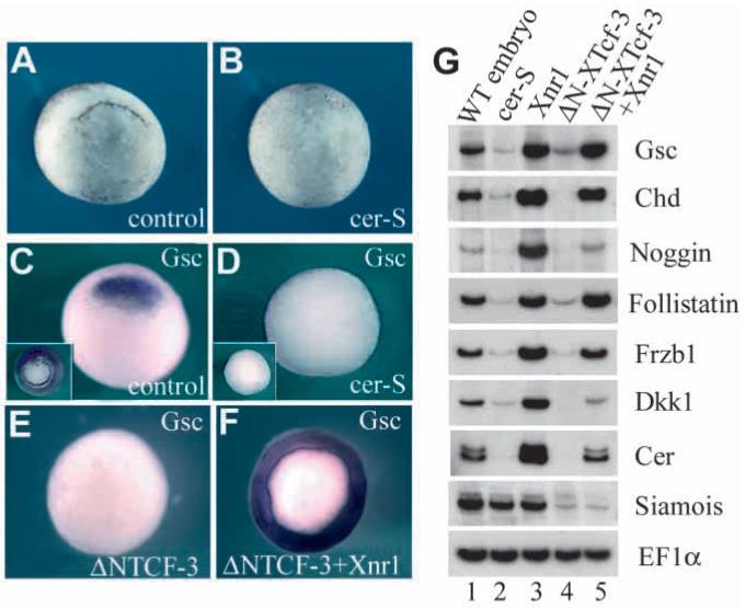
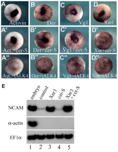
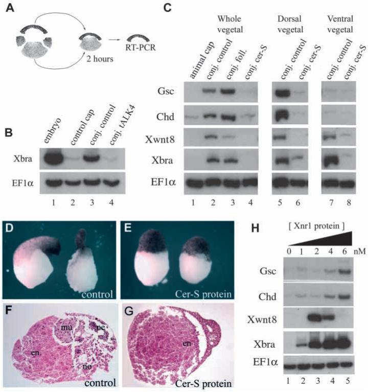
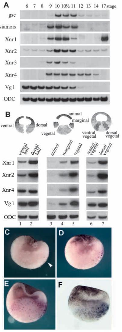
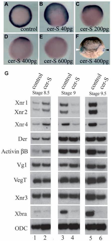
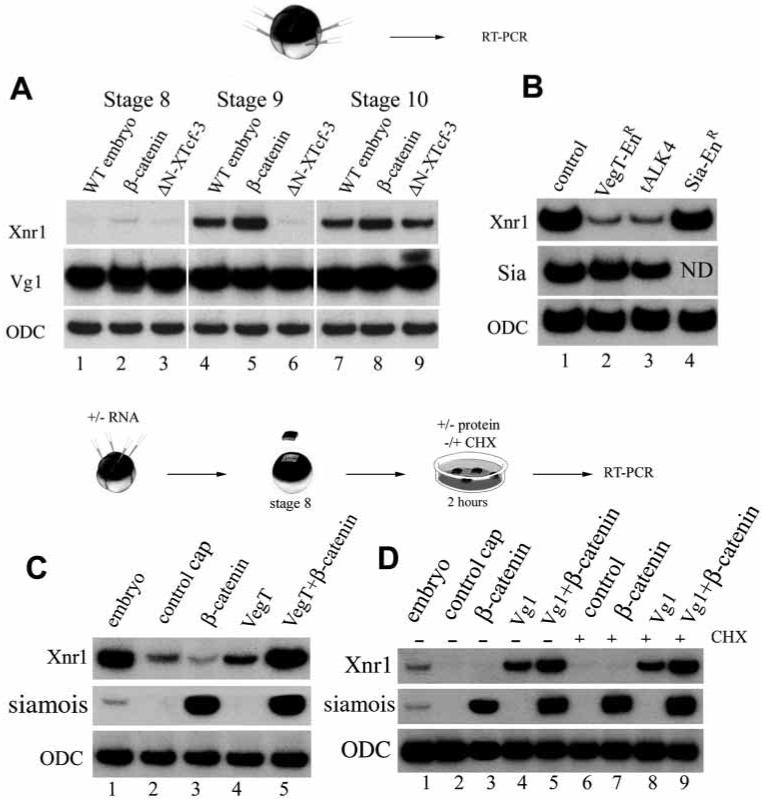
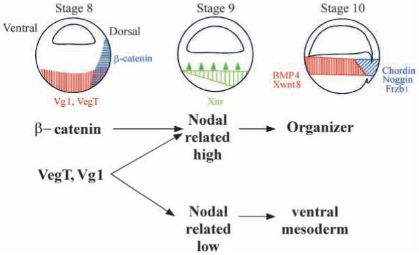
In Xenopus, mesoderm induction by endoderm at the blastula stage is well documented, but the molecular nature of the endogenous inductive signals remains unknown. The carboxy-terminal fragment of Cerberus, designated Cer-S, provides a specific secreted antagonist of mesoderm-inducing Xenopus Nodal-Related (Xnr) factors. Cer-S does not inhibit signalling by other mesoderm inducers such as Activin, Derrière, Vg1 and BMP4, nor by the neural inducer Xnr3. In the present study we show that Cer-S blocks the induction of both dorsal and ventral mesoderm in animal-vegetal Nieuwkoop-type recombinants. During blastula stages Xnr1, Xnr2 and Xnr4 are expressed in a dorsal to ventral gradient in endodermal cells. Dose-response experiments using cer-S mRNA injections support the existence of an endogenous activity gradient of Xnrs. Xnr expression at blastula can be activated by the vegetal determinants VegT and Vg1 acting in synergy with dorsal (beta)-catenin. The data support a modified model for mesoderm induction in Xenopus, in which mesoderm induction is mediated by a gradient of multiple Nodal-related signals released by endoderm at the blastula stage.


Figures

Fig. 1
Fig. 1
Cer-S, a secreted inhibitor of Nodal-related factors, inhibits formation of Spemann’s organizer. (A,B) cer-S mRNA (radial injection of 150 pg into each blastomere at the 4-cell stage) blocks dorsal lip formation. (C,D) gsc expression is blocked by cer-S, even after LiCl treatment that expands gsc expression to the entire mesoderm (insets). (E,F) ΔN-XTcf-3 mRNA (800 pg radially) inhibits goosecoid expression, and co-injection of Xnr1 (50 pg) restores it in the entire marginal zone. (G) RT-PCR analysis of Spemann organizer markers at stage 10. Lane 1: whole embryos. Lane 2: radial injection of cer-S (600 pg total) represses organizer genes. Lane 3: radial injection of Xnr1 mRNA (50 pg) upregulates organizer markers. Lane 4: the β-catenin pathway antagonist ΔN-XTcf-3 (800 pg) inhibits organizer markers, which are rescued by co-injection with 50 pg Xnr1 (lane 5). Xnr1 acts downstream of, or in parallel to, the β-catenin pathway. Siamois (Lemaire et al., 1995) is regulated by the β-catenin pathway (Brannon et al., 1997) independently of Xnr signals.
Fig. 2
Fig. 2
Cer-S inhibits Xnrs but not activin, derrière, Vg1 and Xnr3. Co-injection of cer-S mRNA (150 pg into a single animal blastomere) inhibited ectopic blastopore lip formation by Xnr1 (50 pg, D’), but not by activin (30 pg), derrière (150 pg) or A-Vg1 (50 pg) mRNA (A’-D’). Xnr2 mRNA (150 pg) was also inhibited (data not shown). Although the doses used for each TGF-β mRNA differed, they all were titrated to elicit comparable biological responses. tALK4 (800 pg) mRNA blocked all TGF-β mesoderm inducers tested (A”-D”). The ectopic blastopore lips are seen as a darker area in the injected animal cap region due to the apical constrictions of bottle cells (Lustig et al., 1996). (E) Xnr3 (1.2 ng) is a neural (NCAM) but not mesodermal (α-actin) inducer in microinjected animal caps (lanes 2 and 3). cer-S mRNA (600 pg) does not inhibit Xnr3 activity in animal caps (lanes 4 and 5). EF1α was used as a loading control.
Fig. 3
Fig. 3
The endogenous mesoderm-inducing signals are blocked by Cer-S in Nieuwkoop animal-vegetal conjugates. (A) Experimental design. (B) Microinjection of tALK4 mRNA (500 pg into each animal blastomere at 8-cell stage) blocks the response of animal caps to endogenous mesoderm-inducing signals (compare lanes 3 and 4). Caps were in contact with endoderm for 2 hours and are compared to control animal caps incubated without endoderm (lane 2). EF1α is a control for RNA recovery. (C) Lanes 1-4, Nieuwkoop recombinants of uninjected animal caps with vegetal pole explants injected with follistatin (2 ng) or cer-S (600 pg) mRNA. Note in lane 4 that cer-S blocks dorsal (gsc, chd), ventral (Xwnt-8) and pan-mesodermal (Xbra) markers, whereas in lane 3 follistatin mRNA has only a slight dorsalizing effect (total conjugates n=45, two experiments). This amount of follistatin mRNA was sufficient to abolish the activity of activin mRNA in co-injection assays (not shown). Lane 5, dorsal endoderm (Nieuwkoop center) induces preferentially the organizer markers gsc and chd (n=16, three independent experiments). Lane 7, ventral endoderm induces ventral markers Xwnt-8 and the pan-mesodermal marker Xbra (n=17). Lanes 6 and 8, cer-S mRNA in the endodermal fragment prevents both dorsal and ventral mesoderm inductions (n=15 each). Conjugates were prepared between stage 8 and 8.5 and harvested for RNA after two hours. (D-G) External and histological morphology of vegetal fragments conjugated in the presence of control conditioned medium or of 20 nM Cer-S (Piccolo et al., 1999) protein and cultured until stage 36. Note that sections of the control contain muscle (mu), notochord (no) and some neural tissue (ne), whereas in the protein-treated sample the animal cap remains as atypical epidermis (ae) and endoderm (en) (n=26, three independent experiments). (H) Animal caps treated for two hours with control oocyte conditioned medium (lane 1) or with increasing doses (lanes 2-5) of Xnr1 protein (Piccolo et al., 1999). Increasing concentrations of Xnr1 protein induce first ventral and then dorsal mesodermal markers, producing thresholds after 2 hours in culture.
Fig. 4
Fig. 4
Endogenous Xnrs are expressed at the right time and place to function as mesoderm inducers. (A) Time course of gene expression analyzed by RT-PCR at various developmental stages (Nieuwkoop and Faber, 1994). The mesoderm inducers Xnr1, Xnr2 and Xnr4 start zygotic expression at the same time as Siamois and Xnr3 (which are expressed immediately after midblastula and are direct targets of β-catenin regulation). ODC is used as a loading control. (B) Dissections of embryos at stage 9 showing that Xnr1, Xnr2 and Xnr4 are expressed in the endoderm and at higher levels dorsally than ventrally. Vg1 is expressed uniformly in the vegetal pole. (C-F) Xnr1 in situ hybridizations of blastula stage embryos showing a gradient of expression in endoderm. (C) Stage 8 embryo showing a few nuclei stained in the dorsal vegetal mass (arrowhead). (D) Stage 8.5 blastula embryo in which Xnr1 expression has expanded into neighboring vegetal cells. (E) Stage 9 blastula embryo displaying graded Xnr1 expression throughout the embryonic endoderm. (F) External view of a stage 9 embryo cleared in Murray’s solution in order to visualize Xnr1 staining in the vegetal hemisphere. In this embryo, the ventral side, with its more pigmented animal cap, can be clearly distinguished from the less pigmented dorsal side. Note that Xnr1 expression on the dorsal side is of longer duration, in addition to reaching higher levels than in ventral endoderm.
Fig. 5
Fig. 5
Injections of cer-S mRNA dose-dependently reduce Xbra expression in gastrula embryos. Embryos are injected radially in the vegetal pole at the 4 cell stage, then processed for Xbra in situ staining at stage 10.5. (A) Control uninjected embryo, Xbra is expressed as a mesodermal ring. (B-E) Embryos injected with increasing amounts of cer-S mRNA, showing graded reduction of the Xbra expression domain. (F) Embryos injected vegetally with 400 pg of cer-S mRNA at the 4-cell stage and with lacZ lineage tracer mRNA into blastomere C4 at the 32-cell stage. In this lateral view the white arrowhead indicates lacZ in the ventral side (note that the pigment in the animal cap also marks the ventral side) and the black arrowhead points to the expression of Xbra transcripts on the dorsal side (n=51). (G) RT-PCR analysis of Xenopus embryos injected with 600 pg of cer-S mRNA. RNAs were harvested from uninjected controls or cer-S-injected embryos at one-hour intervals at stages 8.5 (lanes 1, 2), 9.0 (lanes 3, 4) and 9.5 (lanes 5, 6). Xnr1, 2 and 4 transcripts are initially not inhibited by cer-S (lanes 1, 2), but are decreased at later stages (a positive feedback loop for Nodal-related gene expression has been described; Meno et al., 1999). Importantly, the levels of derrière, Vg1, VegT and Xnr3 remained unchanged, and activin βB was only partially decreased. Note that cer-S mRNA inhibited the initial expression of Xbra and that cer-S can inhibit Xbra transcriptional activation even in the presence of derrière, activin and Vg1 transcripts.
Fig. 6
Fig. 6
Zygotic expression of Xnr1 is regulated by β-catenin, VegT and Vg1. (A) Xnr1 is upregulated by β-catenin (200 pg) and inhibited by ΔN-XTcf-3 mRNA (800 pg) at stage 9 (lanes 4-6) in radially injected embryos. At stage 10, however, Xnr1 transcripts are expressed even in the presence of ΔN-XTcf-3; this is consistent with the formation of ventral mesoderm in these ventralized embryos. Maternal Vg1 is not affected. (B) Whole embryos injected radially at 4-cell stage with 800 pg VegT-EnR mRNA (lane 2), 2 ng of tALK4 (lane 3), or 120 pg of Sia-EnR (lane 4) and analyzed by RT-PCR at stage 9. Xnr1 requires VegT activity and TGF-β-like signalling for expression at the blastula stage. Xnr1 expression at blastula is not dependent on Siamois activity since it is not blocked by Sia-EnR mRNA. (C) Animal cap experiments showing that VegT mRNA (200 pg) can weakly induce Xnr1 and synergizes with β-catenin (100 pg) mRNA (lanes 4 and 5). β-catenin mRNA on its own is unable to induce Xnr1 in animal caps (lane 3). (D) Treatment of animal caps with 4 nM Vg1 protein at stage 8 for 2 hours induces Xnr1, and β-catenin (100 pg mRNA) enhances this induction (lanes 2-5). The same response was obtained after blocking protein synthesis with cycloheximide (lanes 6-9), indicating that Xnr1 is a primary response gene to Vg1 protein. Although only results for Xnr1 transcripts are shown, Xnr2 and Xnr4 were also analyzed in all samples of A, C and D with comparable results (data not shown).
Fig. 7
Fig. 7
Model of mesoderm induction at the blastula stage by a dorsal to ventral gradient composed of multiple Nodal-related genes expressed in endoderm. This simplified model concerns the nature of the endodermal signals, but does not address the function of important regulators such as β-catenin and siamois in the marginal zone of the embryo. The model has predictive value as it may help explain how molecules as dissimilar as β-catenin, Vg1, Xnr1 and Xnr2, noggin and chordin mRNAs might rescue dorsal development via a sequential pathway when overexpressed in UV-ventralized embryos.

References

    1. Belo JA, Bouwmeester T, Leyns L, Kertesz N, Gallo M, Follettie M, De Robertis EM. Cerberus-like is a secreted factor with neuralizing activity expressed in the anterior primitive endoderm of the mouse gastrula. Mech. Dev. 1997;68:45–57. - PubMed
    1. Bouwmeester T, Kim SH, Sasai Y, Lu B, De Robertis EM. Cerberus is a head-inducing secreted factor expressed in the anterior endoderm of Spemann’s organizer. Nature. 1996;382:595–601. - PubMed
    1. Boterenbrood EC, Nieuwkoop PD. The formation of the mesoderm in urodelean amphibians. V. Its regional induction by the endoderm. Wilhelm Roux’ Arch. Dev. Biol. 1973;173:319–332. - PubMed
    1. Brannon M, Gomperts M, Sumoy L, Moon RT, Kimelman D. A beta-catenin/XTcf-3 complex binds to the siamois promoter to regulate dorsal axis specification in Xenopus. Genes Dev. 1997;11:2359–2370. - PMC - PubMed
    1. Chang C, Wilson PA, Mathews LS, Hemmati-Brivanlou A. A Xenopus type I activin receptor mediates mesodermal but not neural specification during embryogenesis. Development. 1997;124:827–837. - PubMed

Publication types

MeSH terms