Comparative Study
. 2000 Feb;105(4):497-504.
doi: 10.1172/JCI8541.

Toll-like receptor 4 imparts ligand-specific recognition of bacterial lipopolysaccharide

Affiliations
Comparative Study

Toll-like receptor 4 imparts ligand-specific recognition of bacterial lipopolysaccharide

E Lien et al. J Clin Invest. 2000 Feb.

Abstract

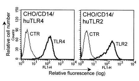
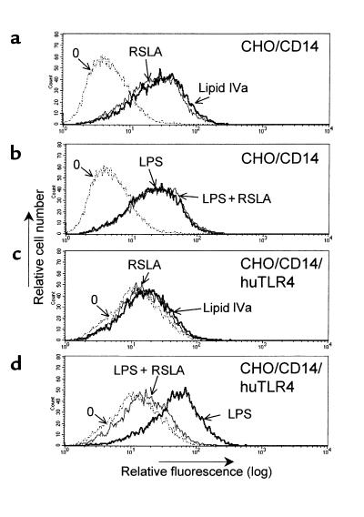
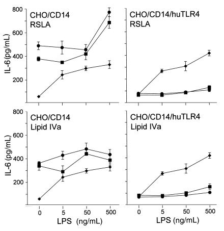
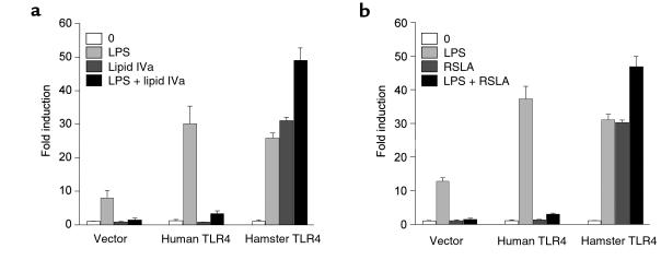
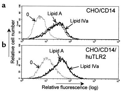
Lipopolysaccharide (LPS) is the main inducer of shock and death in Gram-negative sepsis. Recent evidence suggests that LPS-induced signal transduction begins with CD14-mediated activation of 1 or more Toll-like receptors (TLRs). The lipid A analogues lipid IVa and Rhodobacter sphaeroides lipid A (RSLA) exhibit an uncommon species-specific pharmacology. Both compounds inhibit the effects of LPS in human cells but display LPS-mimetic activity in hamster cells. We transfected human TLR4 or human TLR2 into hamster fibroblasts to determine if either of these LPS signal transducers is responsible for the species-specific pharmacology. RSLA and lipid IVa strongly induced NF-kappaB activity and IL-6 release in Chinese hamster ovary fibroblasts expressing CD14 (CHO/CD14), but these compounds antagonized LPS antagonists in CHO/CD14 fibroblasts that overexpressed human TLR4. No such antagonism occurred in cells overexpressing human TLR2. We cloned TLR4 from hamster macrophages and found that human THP-1 cells expressing the hamster TLR4 responded to lipid IVa as an LPS mimetic, as if they were hamster in origin. Hence, cells heterologously overexpressing TLR4 from different species acquired a pharmacological phenotype with respect to recognition of lipid A substructures that corresponded to the species from which the TLR4 transgene originated. These data suggest that TLR4 is the central lipid A-recognition protein in the LPS receptor complex.


Figures

Figure 1
Figure 1
Expression of human TLR4 and TLR2 transgenes in CHO/CD14 cells. CHO/CD14 cells that were stably transfected with human TLR4 or human TLR2 were labeled with 10 μg/mL of the mAbs HTA125 (TLR4), TL2.1 (TLR2), or a control antibody (CTR, mouse IgG; Sigma-Aldrich), followed by incubation with anti-mouse IgG FITC (Sigma-Aldrich). The cells were subjected to flow cytometry analysis on as described (42). Relative cell number is shown on the y-axis and relative fluorescence is shown on the x-axis.
Figure 2
Figure 2
RSLA blocked LPS-mediated activation in CHO/CD14 reporter cells expressing human TLR4. CHO/CD14 (a and b) and CHO/CD14/huTLR4 (c and d) reporter cells were plated in 24-well dishes. The next day, the cells were exposed to various treatments. (a and c) Cells treated with medium only (stippled lines), RSLA (5 μg/mL; thick lines), or synthetic lipid IVa (5 μg/mL). (b and d) Cells treated with medium only (stippled lines), LPS (0.5 μg/mL; thin lines) or a combination of LPS and RSLA (0.5 and 5 μg/mL, respectively) for 20 hours. After harvesting, the cells were stained for surface CD25 and subjected to flow cytometry analysis. Untreated cells (stippled lines) are indicated by “0”. The x-axis represents relative fluorescence and the y-axis represents relative cell number. One representative experiment out of 5 is shown. Note that although basal immunofluorescence in unstimulated CHO/CD14/TLR4 cells is slightly higher than in CHO/CD14 cells, the ED50 of CHO/CD14/TLR4 to LPS-induced reporter activity has been found to be identical (data not shown).
Figure 3
Figure 3
Lipid IVa and RSLA stimulate the release of the cytokine IL-6 from CD14-expressing hamster fibroblasts but become LPS antagonists when these cells overexpress human TLR4. CHO/CD14 cells and CHO/CD14/huTLR4 cells were allowed to adhere overnight in 24-well dishes. The next day, the cells were exposed to increasing amounts of S. minnesota Re595 LPS in the presence of medium only (diamonds), 0.5 μg/mL lipid IVa or RSLA (squares), or 5 μg/mL of the same compounds (circles) for 10 hours. Supernatants were assayed for IL-6 by bioassay. Data represents mean values of 3 seperate fractions ± SD shown is 1 experiment out of 2 performed.
Figure 4
Figure 4
Reversal of the human phenotype by hamster TLR4 expression: lipid IVa and RSLA activate THP-1 monocytes that overexpress hamster TLR4. THP-1 cells were plated at a density of 2 × 10⁶ cells per well in 6-well dishes, and then transiently transfected with the reporter plasmid pELAM.luc plus either pcDNA3.1 (vector), hToll (human TLR4), or pFLAG-hamTLR4 (hamster TLR4). The next day, the cells were incubated with medium alone (0), LPS (10 ng/mL), lipid IVa (1 μg/mL), or a combination of LPS and lipid IVa (10 ng/mL plus 1 μg/mL, respectively) (a), or in a separate experiment with medium alone (0), LPS (10 ng/mL), RSLA (1 μg/mL), or a combination of LPS and RSLA (10 ng/mL and 1 μg/mL, respectively) (b) for 5 hours. Luciferase activity was measured as described in Methods and plotted as the fold induction of activity compared with vector-transfected, unstimulated controls. The values shown are mean ± SD of triplicate transfections in 1 representative experiment out of 3. Similar results were observed when cells were stimulated with 100 ng/mL LPS and a 10-fold excess of inhibitor (data not shown).
Figure 5
Figure 5
Expression of human TLR2 in CHO/CD14 cells does not change the responses to lipid IVa and synthetic lipid A. Untransfected CHO/CD14 reporter cells (a) or CHO/CD14 reporter cells stably transfected with human TLR2 (29) (b) were stimulated with synthetic lipid A (0.5 μg/mL) as a positive control, or with lipid IVa (0.5 μg/mL) as indicated. After 20 hours, the cells were harvested and stained for surface CD25 and then subjected to flow cytometry analysis. Untreated cells (stippled lines) are indicated by “0”. The x-axis represents relative fluorescence, and the y-axis represents relative cell number. Results shown are from 1 of 2 experiments performed. Similar results were observed in RSLA-exposed cells and with CHO/CD14/moTLR2 cells treated with either lipid IVa or RSLA.

References

    1. Sands KE, et al. Epidemiology of sepsis syndrome in 8 academic medical centers. Academic Medical Center Consortium Sepsis Project Working Group. JAMA. 1997;278:234–240. - PubMed
    1. Raetz CR, et al. Gram-negative endotoxin: an extraordinary lipid with profound effects on eukaryotic signal transduction. FASEB J. 1991;5:2652–2660. - PubMed
    1. Fenton MJ, Golenbock DT. LPS-binding proteins and receptors. J Leukoc Biol. 1998;64:25–32. - PubMed
    1. Kitchens RL, Munford RS. Enzymatically deacylated lipopolysaccharide (LPS) can antagonize LPS at multiple sites in the LPS recognition pathway. J Biol Chem. 1995;270:9904–9910. - PubMed
    1. Kitchens RL, Ulevitch RJ, Munford RS. Lipopolysaccharide (LPS) partial structures inhibit responses to LPS in a human macrophage cell line without inhibiting LPS uptake by a CD14-mediated pathway. J Exp Med. 1992;176:485–494. - PMC - PubMed

Publication types

MeSH terms