. 2000 Feb;105(4):533-9.
doi: 10.1172/JCI8625.

Pretreatment with DNA-damaging agents permits selective killing of checkpoint-deficient cells by microtubule-active drugs

Affiliations

Pretreatment with DNA-damaging agents permits selective killing of checkpoint-deficient cells by microtubule-active drugs

M V Blagosklonny et al. J Clin Invest. 2000 Feb.

Abstract

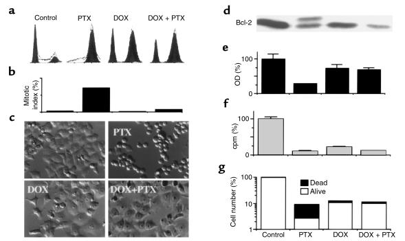
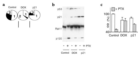
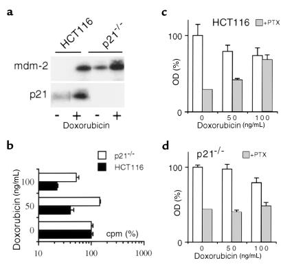
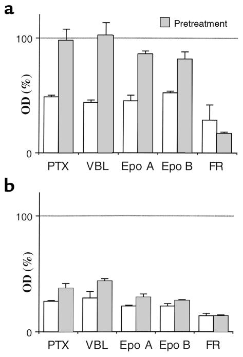
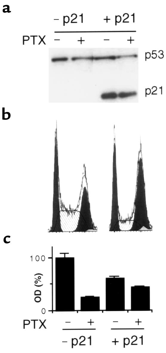
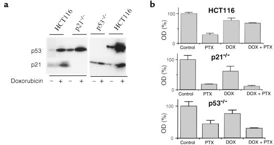
Cell-cycle checkpoint mechanisms, including the p53- and p21-dependent G(2) arrest that follows DNA damage, are often lost during tumorigenesis. We have exploited the ability of DNA-damaging drugs to elicit this checkpoint, and we show here that such treatment allows microtubule drugs, which cause cell death secondary to mitotic arrest, to kill checkpoint-deficient tumor cells while sparing checkpoint-competent cells. Low doses of the DNA-damaging drug doxorubicin cause predominantly G(2) arrest without killing HCT116 cells that harbor wt p53. Doxorubicin treatment prevented mitotic arrest, Bcl-2 phosphorylation, and cell death caused by paclitaxel, epothilones, and vinblastine. In contrast, doxorubicin enhanced cytotoxicity of FR901228, an agent that does not affect microtubules. Low doses of doxorubicin did not arrest p21-deficient clones of HCT116 cells and did not protect these cells from cytotoxicity caused by microtubule drugs, but cells in which p21 expression was restored enjoyed partial protection under these conditions. Moreover, in p53-deficient clones of HCT116 cells doxorubicin did not induce either p53 or p21 and provided no protection against paclitaxel-induced cytotoxicity. Therefore, (a) p53-dependent p21 induction caused by doxorubicin protects from microtubule drug-induced cytotoxicity, and (b) pretreatment with cytostatic doses of DNA-damaging drugs before treatment with microtubule drugs results in selective cytotoxicity to cancer cells with defective p53/p21-dependent checkpoint.


Figures

Figure 1
Figure 1
Doxorubicin-induced G2 arrest versus paclitaxel-induced mitotic arrest, Bcl-2 phosphorylation and cell death. HCT116 cells were incubated with 100 ng/mL doxorubicin (DOX) or left untreated. After 12–16 hours, 100 ng/mL paclitaxel (PTX) was added, if indicated. Pretreated cells continued in doxorubicin until harvest. (a) Cell-cycle distribution measured by FACS analysis was performed 24 hours after the addition of PTX. (b) Mitotic index was measured by DAPI staining after 24 hours after the addition of PTX. (c) Cells, treated as indicated, were photographed 16 hours after the addition of PTX. (d) Bcl-2 protein phosphorylation was demonstrated by immunoblot 12 hours after the addition of PTX. (e) Number of live cells, as measured by MTT assay, 2 days after the addition of PTX. (f) DNA replication, as measured by [3H]thymidine incorporation, 16 hours after the addition of PTX. (g) The number of dead and live cells was counted with trypan blue staining 3 days after the addition of PTX. Open bars represent live cells (excluded trypan blue); filled bars represent dead cells (stained with trypan blue).
Figure 2
Figure 2
Comparison of DOX- and p21-mediated cytoprotection. (a) HCT116 cells were treated with 100 ng/mL doxorubicin (DOX) or infected with 10 moi Ad-p21 (p21) for 36 hours, and then cell-cycle analysis was performed as described in Methods. The pie chart represents the percent of cells in each phase of the cell cycle: 1 = G1 phase; 2 = G2 phase; solid area = S phase. Control adenovirus, Ad-LacZ, did not change cell-cycle distribution. (b) HCT116 cells were pretreated with 100 ng/mL DOX for 16 hours or 10 moi Ad-p21 (p21) for 24 hours. A plus sign indicates that cells were treated with 100 ng/mL PTX for 20 hours; a minus sign indicates that they were left untreated. Cells were lysed after 20 hours, and p53, p21, Raf-1, and p120 were measured by immunoblot. (c) HCT116 cells were pretreated as already described here and then treated with 100 ng/mL PTX. After 48 hours the MTT assay was performed.
Figure 3
Figure 3
Doxorubicin does not protect p21–/– cells. (a) HCT116 and p21–/– cells were incubated with 100 ng/mL doxorubicin for 24 hours and then assayed for mdm-2 and p21 proteins by immunoblot analysis. (b) HCT116 and p21–/– cells were incubated with the indicated concentrations of doxorubicin for 24 hours, and then [3H]thymidine incorporation was measured. (c and d) HCT116 (c) and p21–/– cells (d) were pretreated with indicated dose of doxorubicin for 12 hours, and then cells were treated with 100 ng/mL PTX (gray bars) or left untreated (open bars). MTT test was performed after 2 days.
Figure 4
Figure 4
Comparison of PTX with other microtubule-active drugs. HCT116 cells (a) or p21–/– cells (b) were pretreated with 100 ng/mL doxorubicin or left without pretreatment. After 16 hours, cells were treated with 100 ng/mL of indicated drug. MTT assay was performed after 2 days. Results represent percent of untreated cells or DOX-pretreated cells, respectively. Epo, epothilone. VBL, vinblastine.
Figure 5
Figure 5
Protection by p21 against PTX in p21–/–. (a) Cells were infected with 10 moi Ad-p21, and after 24 hours, treated with 100 ng/mL PTX for 20 hours as indicated. Cells were lysed, and immunoblot for p53 and p21 was performed. (b) Cells were infected with 10 moi Ad-p21, and after 24 hours, cell-cycle analysis was performed. No doxorubicin or paclitaxel was added in this experiment. (c). Cells were infected with 10 moi Ad-p21, and after 24 hours, treated with 100 ng/mL PTX as indicated. MTT assays were performed after 2 days.
Figure 6
Figure 6
Doxorubicin fails to protect p53–/– cells. (a) Effects of doxorubicin on p53 and p21 proteins in HCT116, p21–/–, and p53–/– cells. Cells were incubated with (+) or without (–) 100 ng/mL doxorubicin for 16 hours and then assayed by immunoblot analysis. (b) Cells were pretreated with 100 ng/mL doxorubicin as indicated (DOX) and continued with or without 100 ng/mL paclitaxel (PTX). Cell survival was measured by MTT assay 2 days after PTX addition.

References

    1. Horwitz SB. Mechanism of action of taxol. Trends Pharmacol Sci. 1992;13:134–136. - PubMed
    1. Rowinsky EK, Donehower RC. Paclitaxel (Taxol) N Engl J Med. 1995;332:1004–1014. - PubMed
    1. Kastan MB, Onyekwere O, Sidransky D, Vogelstein B, Craig RW. Participation of p53 protein in the cellular response to DNA damage. Cancer Res. 1991;51:6304–6311. - PubMed
    1. El-Deiry WS, et al. WAF1, a potential mediator of p53 tumor suppression. Cell. 1993;75:817–825. - PubMed
    1. Brugarolas J, et al. Radiation-induced cell cycle arrest compromised by p21 deficiency. Nature. 1995;377:552–557. - PubMed