. 2000 Feb 21;148(4):703-14.
doi: 10.1083/jcb.148.4.703.

Debcl, a proapoptotic Bcl-2 homologue, is a component of the Drosophila melanogaster cell death machinery

Affiliations

Debcl, a proapoptotic Bcl-2 homologue, is a component of the Drosophila melanogaster cell death machinery

P A Colussi et al. J Cell Biol. .

Abstract

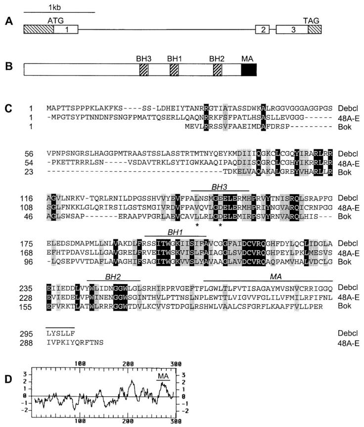
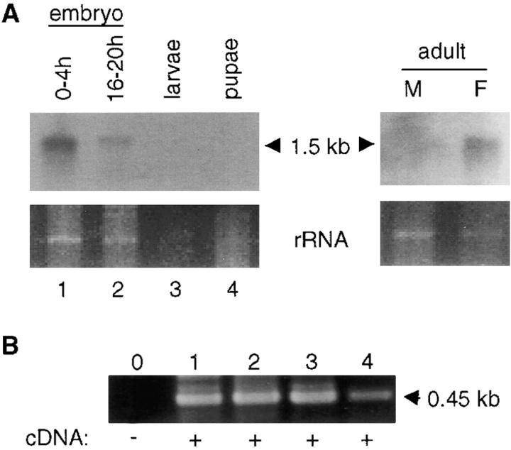
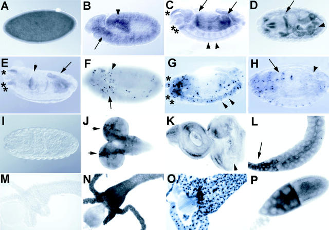
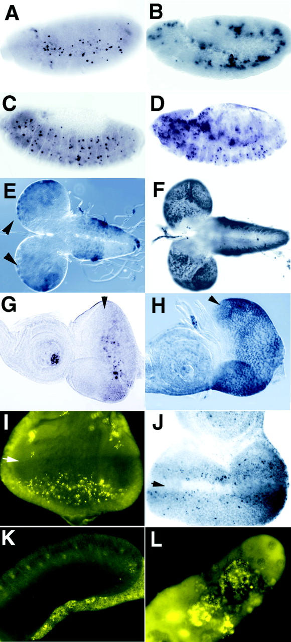
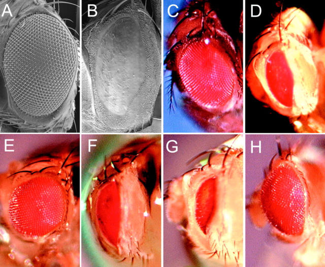
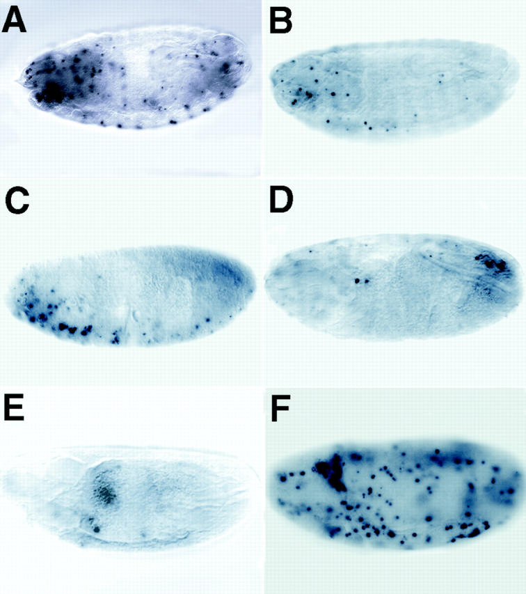
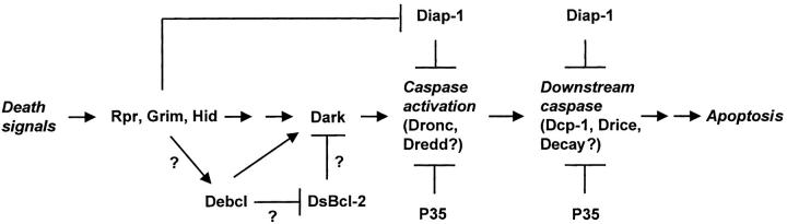
Bcl-2 family of proteins are key regulators of apoptosis. Both proapoptotic and antiapoptotic members of this family are found in mammalian cells, but no such proteins have been described in insects. Here, we report the identification and characterization of Debcl, the first Bcl-2 homologue in Drosophila melanogaster. Structurally, Debcl is similar to Bax-like proapoptotic Bcl-2 family members. Ectopic expression of Debcl in cultured cells and in transgenic flies causes apoptosis, which is inhibited by coexpression of the baculovirus caspase inhibitor P35, indicating that Debcl is a proapoptotic protein that functions in a caspase-dependent manner. debcl expression correlates with developmental cell death in specific Drosophila tissues. We also show that debcl genetically interacts with diap1 and dark, and that debcl-mediated apoptosis is not affected by gene dosage of rpr, hid, and grim. Biochemically, Debcl can interact with several mammalian and viral prosurvival Bcl-2 family members, but not with the proapoptotic members, suggesting that it may regulate apoptosis by antagonizing prosurvival Bcl-2 proteins. RNA interference studies indicate that Debcl is required for developmental apoptosis in Drosophila embryos. These results suggest that the main components of the mammalian apoptosis machinery are conserved in insects.


Figures

Figure 1
Figure 1
Debcl is a Bcl-2–like protein. A, Genomic structure of the debcl gene at 42E-43A. The noncoding regions of the exons are shown as hatched boxes. B, Debcl protein structure. The relative positions of the three BH domains (BH1, BH2, and BH3) and a membrane anchor (MA) are shown. C, An alignment of the Debcl sequence with Bok and 48A-E Drosophila Bcl-2 homologue. The sequence of the 48A-E homologue was obtained from a partial cDNA sequence isolated by us and the genomic sequence in the data base. The protein sequence of this clone is likely to be incomplete at the NH2 terminus. Residues identical in all three proteins are shown in black boxes and those similar shown in gray boxes. The positions of the two residues in the BH3 domain of Debcl, which were mutated in functional studies in Fig. 5 A, are indicated by an asterisk. D, A Kyte-Doolittle plot of the Debcl protein showing the putative MA region.
Figure 2
Figure 2
debcl mRNA expression in Drosophila. A, Northern blot of poly A+-enriched RNA isolated from various developmental stages and adult flies. debcl transcript is detected as a single, ∼1.5-kb band in most samples examined. The lower panels depict portions of the ethidium bromide-stained gels corresponding to the residual ribosomal RNA bands before transfer to membrane. B, RT-PCR analysis of debcl expression. After reverse transcription of RNA from various stages of Drosophila development, PCR was carried out for 30 cycles using debcl-specific primers that generate a 450-bp product. Lanes 1–4 in B correspond to lanes 1–4 in A. Note that all samples express debcl transcript.
Figure 3
Figure 3
In situ analysis of debcl expression during development. A debcl antisense RNA probe labeled with digoxygenin was used to detect debcl expression in situ. A, Uniform staining is evident in a stage 5 cellularized embryo. B, In germ band extended embryo (stage 11), staining is evident in the anterior (arrow) proctodeum and posterior midgut (arrowhead), which are regions that show higher levels of TUNEL positivity (F). C, A lateral view of a germ band retracted embryo (stage 14) showing staining in the gut, particularly in the anterior and posterior midgut (arrows) and staining in the head corresponding to tissues of the clypeolabrum (*) and of the pharynx (**). Staining is also observed in a segmental reiterated pattern (examples indicated by arrowheads), that may correspond to cells in the central and peripheral nervous system, which show positive TUNEL at this stage (G). D, A dorsal view of a stage 16 embryo showing staining in regions in the head and gut (arrow indicates a strong stripe of staining that occurs in the foregut–midgut junction). E, A lateral view of an embryo at stage 16, showing staining in regions of the gut (arrowhead indicates the foregut–midgut junction and arrow indicates the hindgut), and in tissues of the clypeolabrum (*) and pharynx (**). F, TUNEL of an embryo at stage 11, showing a higher level of TUNEL positive cells in the region of the anterior midgut (arrow) and the proctodeum, posterior midgut (arrowhead). G, TUNEL of a stage 14 embryo, showing TUNEL positive cells in a segmentally reiterated pattern in cells of the nervous system (examples indicated by arrowheads) and in the region of the clypeolabrum (*) and pharynx (**). H, TUNEL on a stage 16 embryo showing higher numbers of TUNEL positive cells in the gut (midgut indicated by the arrow, and hindgut indicated by the arrowhead), and in head. I, A stage 16 embryo hybridized with a control sense probe. J, Antisense probe on third instar larval brain lobes showing stronger staining in rows of cells in the region of the outer proliferative center of the brain hemispheres (indicated by arrows), a region that also labels with TUNEL (see Fig. 4 E). K, Antisense probe on a third instar larval eye-antennal disc showing weak staining. The arrowhead indicates the morphogenetic furrow, after which higher levels of staining are observed in some cells corresponding with the region where TUNEL positive cells are observed (see Fig. 4 G). L, Antisense probe on third instar larval salivary glands showing positivity in the duct (arrow). M, Sense control probe on third instar larval gut showing no staining. Also, sense controls on other larval tissues and adult ovaries showed no staining (data not shown). N, Antisense probe on late third instar larval gut showing high levels of staining. O, TUNEL of a late third instar larval gut showing most cells are positive at this stage. P, Antisense probe on ovaries showing high levels of debcl expression in the nurse cells (on the left) and in the oocyte (right) of stage 10a egg chambers. Lower levels of staining are observed subsequent to stage 10 (not shown).
Figure 4
Figure 4
Debcl induces cell death in vivo. Homozygous flies containing debcl under control of the UAS-GAL4 promoter were crossed to various GAL4 drivers and the effect on cell death examined by TUNEL or acridine orange staining. B, D, F, and H represent samples from hsp70-GAL4 × UAS-debcl after heat shock induction. Samples were heat-shocked for 1 h and allowed to recover for 1 h (A and B) or for 3 h (C–H) before fixation and staining. A, A wild-type stage 11 embryo after heat shock, showing a normal pattern of TUNEL. B, A hsp70-GAL4 × UAS-debcl stage 11 embryo after heat shock-induced expression, showing an increase in TUNEL positive cells relative to A. C, A wild-type stage 13 embryo after heat shock showing a normal pattern of TUNEL. D, A hsp70-GAL4 × UAS-debcl stage 13 embryo after heat shock showing an increase in TUNEL positive cells relative to C. Strong TUNEL positive cells are observed in the gut (out of the plane of focus). E, A wild-type 3rd instar larval brain lobe (dorsal view) after heat shock showing low levels of TUNEL staining cells in the brain hemispheres (arrowheads) and in the ventral ganglion. F, A hsp70-GAL4 × UAS-debcl 3rd instar larval brain lobe (dorsal view) after heat shock-induced expression showing an increase in TUNEL positivity relative to E. Note that most of the TUNEL positive cells in the ventral ganglion are out of the plane of focus but extend all the way to the posterior end. G, A wild-type 3rd instar larval eye-antennal disc after heat shock showing only a few TUNEL positive cells. The arrowhead indicates the morphogenetic furrow (also in H, I, and J) after which there are a cluster of TUNEL positive cells. H, A hsp70-GAL4 × UAS-debcl 3rd instar larval eye-antennal disc after heat shock expression showing a large increase in TUNEL positive cells relative to G. I, Acridine orange staining of an eye disc from GMR-GAL4 × UAS-debcl flies, which results in expression in the posterior region of the eye disc, showing an increase in apoptotic cells in the posterior region. Acridine orange staining of control discs was similar to TUNEL labeling (not shown). J, TUNEL staining of an eye disc from eyeless-GAL4 × UAS-debcl flies, which results in expression throughout the eye disc during 2nd instar larval development, and is strong in the anterior region in 3rd instar larvae showing an increase in TUNEL positive cells anterior to the morphogenetic furrow (arrow). K, Acridine orange staining of a 3rd instar larval salivary gland showing essentially no staining, even after long exposure. L, Acridine orange staining of a 3rd instar larval salivary gland from a 109-88-GAL4 × UAS-debcl, which results in strong expression in the embryonic and larval salivary glands (not shown), showing that there is strong staining of the large polyploid nuclei.
Figure 6
Figure 6
Genetic interactions of GMR-p35, the Df(3L)H99 genes, diap1 and dark with GMR-GAL4; UAS-debcl#26. The effects of GMR-p35 and reducing the dosage of the Df(3L)H99 genes, diap1 and dark genes on the eye phenotype of heterozygous GMR-GAL4; UAS-debcl#26 flies, were examined after crossing GMR-GAL4/CyO; UAS-debcl#26/TM6B flies to the relevant stocks. A, Scanning electron micrograph of a wild-type adult eye (Canton S). B, Scanning electron micrograph of GMR-GAL4; UAS-debcl#26 adult eye, showing severe ablation of the eye. C, Photograph of a wild-type eye (Canton S). D, Photograph of GMR-GAL4; UAS-debcl#26 adult eye, showing severe ablation of the eye and patches of reduced pigmentation. E, Photograph of GMR-GAL4; UAS-debcl#26/GMR-p35 adult eye, showing strong suppression of the ablated eye phenotype. F, Photograph of GMR-GAL4; UAS-debcl#26/Df(3L)H99 (removing rpr, hid, and grim) adult eye, showing little effect on the ablated eye phenotype. G, Photograph of GMR-GAL4; UAS-debcl#26/Df(3L)brm11 (removing diap1) adult eye, showing enhancement of the ablated eye phenotype. Similar results were obtained using another diap1 deficiency, (Df(3L)stf-13). H, Photograph of GMR-GAL4/darkCD8 (hypomorphic allele); UAS-debcl#26 showing suppression.
Figure 8
Figure 8
debcl is required for developmental cell death in embryos. RNAi was used to ablate debcl function in embryos. Precellularized embryos were injected with double-stranded debcl RNA and aged to stage 16 before fixation and TUNEL labeling. A, An uninjected control embryo showing the normal pattern of TUNEL labeling. B–E, Typical examples of injected embryos from the debcl RNAi experiment showing that the number of TUNEL positive cells is dramatically reduced compared with the control (A; see Fig. 3 H). F, A buffer-injected control shows that the injection procedure does not inhibit apoptosis, but instead an increase in TUNEL positive cells is observed (compare A and F).
Figure 9
Figure 9
Possible position of Debcl in the Drosophila cell death pathway leading to caspase activation. Several genetic and biochemical studies have established the possible hierarchy between various components of the Drosophila cell death machinery (reviewed in Abrams 1999). The studies described in this paper place Debcl upstream of the P35-inhibitable, Dark-mediated caspase activation pathway. Debcl may lie downstream of the proteins of the H99 complex (Reaper, Grim, and Hid). However, further experiments are required to firmly place Debcl and H99 in the same genetic pathway. Similar to mammalian death pathways, Debcl may function by antagonizing a yet undiscovered prosurvival Bcl-2–like protein(s) (shown as DsBcl-2) in Drosophila.

Comment in

References

    1. Abrams J.M. An emerging blueprint for apoptosis in Drosophila . Trends Cell Biol. 1999;9:435–440. - PubMed
    1. Abrams J.M., White K., Fessler L.I., Steller H. Programmed cell death during Drosophila embryogenesis. Development. 1993;117:29–43. - PubMed
    1. Adams J.M., Cory S. The Bcl-2 protein familyarbiters of cell survival. Science. 1998;281:1322–1326. - PubMed
    1. Antonsson B., Conti F., Ciavatta A., Montessuit S., Lewis S., Martinou I., Bernasconi L., Bernard A., Mermod J.J., Mazzei G. Inhibition of Bax channel-forming activity by Bcl-2. Science. 1997;277:370–372. - PubMed
    1. Bhat M.A., Izaddoost S., Lu Y., Cho K.O., Choi K.W., Bellen H.J. Discs Lost, a novel multi-PDZ domain protein, establishes and maintains epithelial polarity. Cell. 1999;96:833–845. - PubMed

Publication types

MeSH terms

Associated data