. 2000 Mar;74(6):2567-83.
doi: 10.1128/jvi.74.6.2567-2583.2000.

Efficient gene transfer into human CD34(+) cells by a retargeted adenovirus vector

Affiliations

Efficient gene transfer into human CD34(+) cells by a retargeted adenovirus vector

D M Shayakhmetov et al. J Virol. 2000 Mar.

Abstract

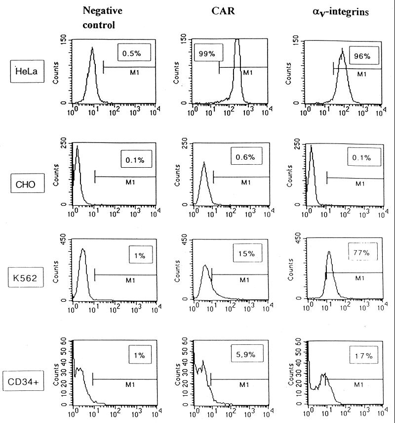
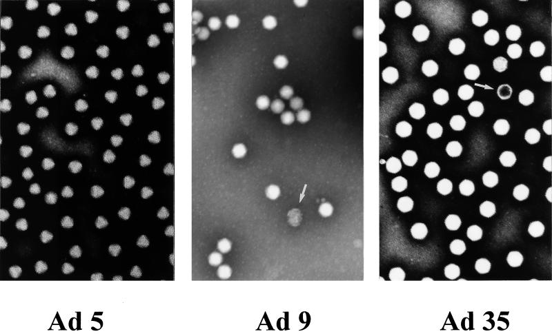
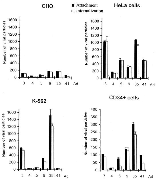
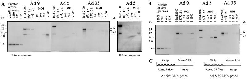
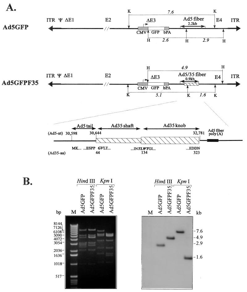
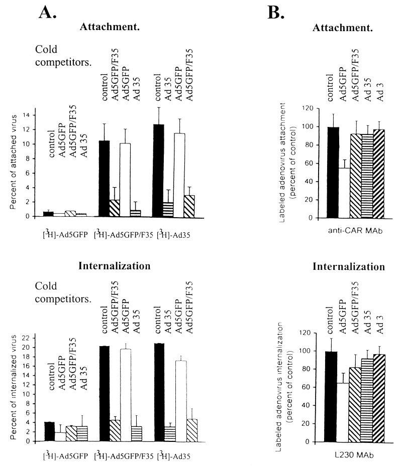
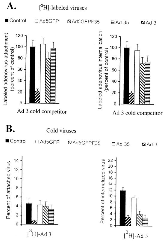
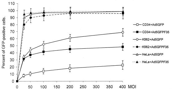
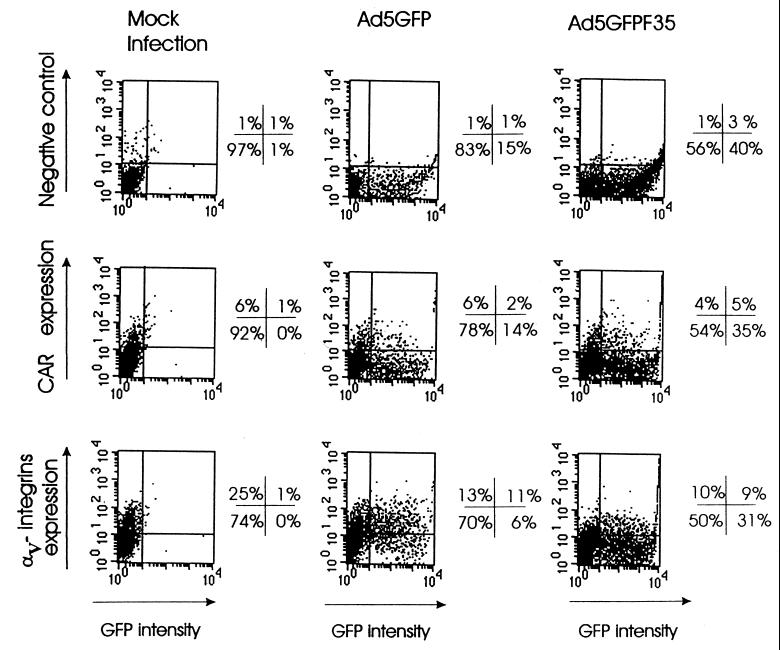
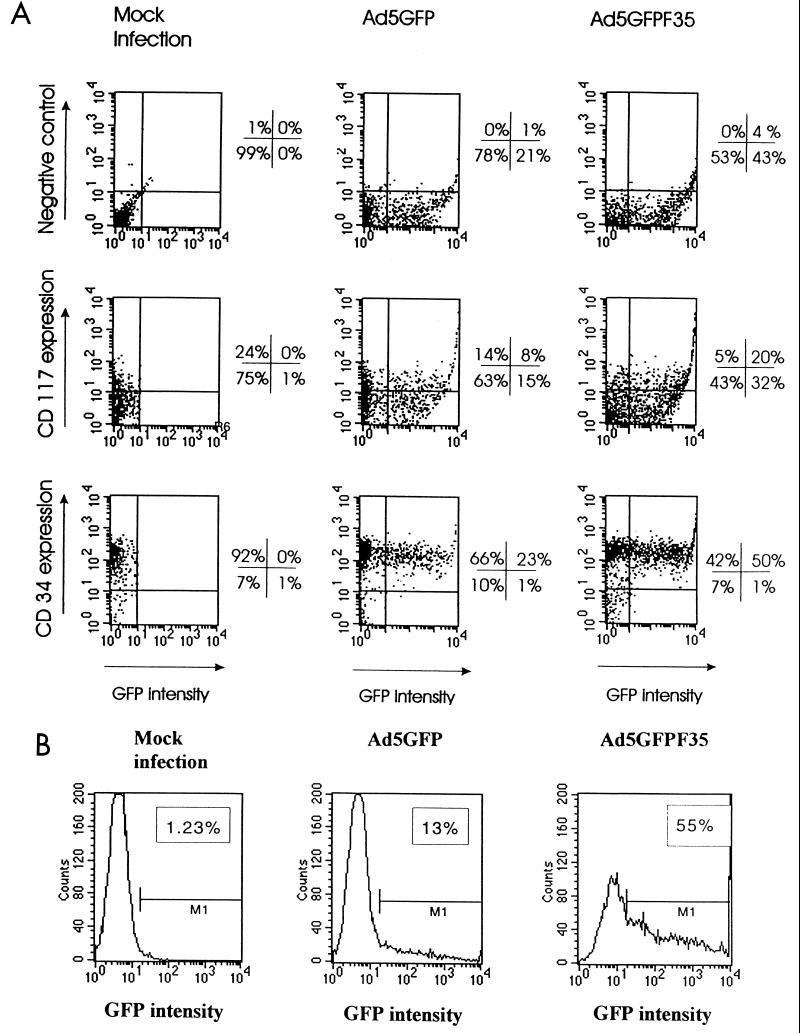
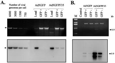
Efficient infection with adenovirus (Ad) vectors based on serotype 5 (Ad5) requires the presence of coxsackievirus-adenovirus receptors (CAR) and alpha(v) integrins on cells. The paucity of these cellular receptors is thought to be a limiting factor for Ad gene transfer into hematopoietic stem cells. In a systematic approach, we screened different Ad serotypes for interaction with noncycling human CD34(+) cells and K562 cells on the level of virus attachment, internalization, and replication. From these studies, serotype 35 emerged as the variant with the highest tropism for CD34(+) cells. A chimeric vector (Ad5GFP/F35) was generated which contained the short-shafted Ad35 fiber incorporated into an Ad5 capsid. This substitution was sufficient to transplant all infection properties from Ad35 to the chimeric vector. The retargeted, chimeric vector attached to a receptor different from CAR and entered cells by an alpha(v) integrin-independent pathway. In transduction studies, Ad5GFP/F35 expressed green fluorescent protein (GFP) in 54% of CD34(+) cells. In comparison, the standard Ad5GFP vector conferred GFP expression to only 25% of CD34(+) cells. Importantly, Ad5GFP transduction, but not Ad5GFP/F35, was restricted to a specific subset of CD34(+) cells expressing alpha(v) integrins. The actual transduction efficiency was even higher than 50% because Ad5GFP/F35 viral genomes were found in GFP-negative CD34(+) cell fractions, indicating that the cytomegalovirus promoter used for transgene expression was not active in all transduced cells. The chimeric vector allowed for gene transfer into a broader spectrum of CD34(+) cells, including subsets with potential stem cell capacity. Fifty-five percent of CD34(+) c-Kit(+) cells expressed GFP after infection with Ad5GFP/F35, whereas only 13% of CD34(+) c-Kit(+) cells were GFP positive after infection with Ad5GFP. These findings represent the basis for studies aimed toward stable gene transfer into hematopoietic stem cells.


Figures

FIG. 1
FIG. 1
Expression of CAR and αv integrins on test cells. For flow cytometry analysis, HeLa, CHO, K562, and CD34+ cells were incubated with an anti-CAR (RmcB; 1:400 dilution) or anti-αv integrin (L230; 1:30 dilution) MAb. As a negative control, cells were incubated with an irrelevant mouse MAb (anti-BrdU; 1:100 dilution). The binding of primary antibodies was developed with anti-mouse IgG-FITC conjugates (1:100 dilution). Data shown represent the average results of quadruplicate analyses performed on 104 cells.
FIG. 2
FIG. 2
EM analysis of Ad particles. Purified particles from Ad5, Ad9, and Ad35 were negative contrast stained and analyzed at an original magnification of ×85,000. Defective particles are highlighted by arrows.
FIG. 3
FIG. 3
Analysis of attachment and internalization of different serotypes to CHO, HeLa, K562, and CD34+ cells. Equal amounts of [3H]thymidine-labeled virions of Ad serotypes 3, 4, 5, 9, 35, and 41 (measured by OD260 and equivalent to an MOI of 400 PFU per cell for Ad5) were incubated for 1 h on ice as described in Materials and Methods. Cells were then washed, and the number of labeled virions bound per cell was determined. For internalization studies, viruses were first allowed to attach to cells for 1 h on ice, and then unbound viral particles were washed out. Cells were then incubated at 37°C for 30 min, treated with trypsin-EDTA, and washed to remove uninternalized viral particles. The data were obtained from two to four independent experiments performed in triplicate. Note the different scales on the y axes for CD34+ cells.
FIG. 4
FIG. 4
Analysis of viral replication in K562 and CD34+ cells by Southern blot analysis of methylated viral DNA. Replication studies were performed with 105 K562 (A) or CD34+ (B) cells infected with methylated Ad5, Ad9, or Ad35. The lanes labeled “load” represent DNA that was extracted from the medium-cell mixture immediately after adding the indicated viral dose to cells. The intensities of bands corresponding to methylated and unmethylated viral DNA indicate that ∼85% of the input virus was methylated. To quantify adsorption and internalization, DNA analysis was performed after prior incubation of virus with cells at 0°C (adsorption) or 37°C (internalization). For dose-dependent replication studies, the indicated viral dose (expressed as the number of genomes per cell) was added, and cellular genomic DNA together with viral DNA was extracted 16 or 36 h postinfection for K562 and CD34+ cells, respectively. Identical amounts of sample DNA were analyzed by Southern blotting. For quantification purposes, Ad9 replication was analyzed together with Ad5, using an Ad5/9 chimeric probe that hybridizes with DNA of both serotypes (C). The analysis of Ad5 versus Ad35 replication was performed with the corresponding Ad5/35 chimeric probe. Since separate hybridizations with both Ad5/35 and Ad5/9 probes gave identical signal intensities for Ad5 DNA, only one panel is shown for Ad5 replication in test cells. To produce distinguishable fragments specific for the methylated or nonmethylated status of viral genomes, Ad5 DNA was digested with XhoI, while Ad9 and Ad35 DNA was digested with XhoI and HindIII. The bands specific for methylated (not replicated) viral DNA were ∼12 kb for Ad9, 35 kb for Ad5, and ∼12 kb for Ad35. The fragments specific for nonmethylated DNA were 5.8 kb for Ad9, 6.1 kb for Ad5, and 9.5 kb for Ad35. Chimeric Ad5/9 and Ad5/35 DNA fragments (1.8 kb) were used as quantification standards and applied onto gels together with digested viral or cellular DNA (shown on the left). Hybridized blots were subjected to quantitative phosphorimager analysis or exposed to X-ray films for 12 h. Lanes representing Ad5 replication data for K562 cells were also exposed for 48 h (A, left panel).
FIG. 5
FIG. 5
Structure of Ad5GFP and chimeric Ad5GFP/F35 vectors. (A) Schematic diagram of the original E1/E3-deleted Ad5-based vector with GFP expression cassette inserted into the E3 region (Ad5GFP) and the chimeric vector Ad5GFP/F35 containing the Ad5/35 fiber gene. The 2.2-kb Ad5 fiber gene was replaced by a 0.9-kb chimeric fiber gene encoding the short shaft and knob of Ad35 by a technique that involved PCR cloning and recombination in E. coli. KpnI (K) and HindIII (H) sites localized within or around the fiber genes are indicated. The lower panel shows the detailed structure of the chimeric fiber region. The Ad5 fiber tail (amino acids [aa] 1 to 44) were joined in frame to the Ad35 fiber shaft starting from its first two amino acids (GV), which are conserved among many serotypes. A conserved stretch of amino acids, TLWT, marks the boundary between the last β sheet of Ad35 shaft and the globular knob. The Ad35 fiber chain termination codon is followed by the Ad5 fiber polyadenylation signal. The region of Ad5GFP/F35 encoding for chimeric fiber was completely sequenced with Ad5-specific primers (see Materials and Methods). ITR, inverted terminal repeat; bPA, bovine growth hormone polyadenylation signal. (B) Restriction analysis of viral genomes. Viral DNA was isolated from purified Ad5GFP and Ad5GFP/F35 particles as described elsewhere (41). One microgram of DNA was digested with HindIII or KpnI and separated in ethidium bromide-stained agarose gels (left) which were subsequently blotted and analyzed by Southern blot with an Ad5 E4-specific probe (nt 32775 to 33651) (right). Specific patterns designating the correct structure for both viral vectors were detected. The HindIII fragments specific for Ad5GFP and Ad5GFP/F35 were 2.9 and 4.9 kb, respectively. The KpnI fragment that confirmed the correct Ad5GFP/F35 structure was 1.6 kb, compared to a 7.6-kb Ad5GFP fragment. M, 1-kb ladder (Gibco-BRL, Grand Island, N.Y.).
FIG. 6
FIG. 6
Cross-competition for attachment and internalization of labeled Ad5GFP, Ad35, and chimeric Ad5GFP/F35 virions with unlabeled viruses and with anti-CAR or anti-αv integrin MAb. (A) For attachment studies, 105 K562 cells were preincubated with a 100-fold excess of unlabeled competitor virus (cold competitors) at 4°C for 1 h; then equal amounts of [3H]Ad5GFP, [3H]Ad5GFP/F35, or [3H]Ad35, at a dose equivalent to an MOI of 100 PFU per cell determined for Ad5GFP, were added to cells, which were incubated at 4°C for 1 h. Cells were then washed with ice-cold PBS and pelleted, and the percentage of attached virus (cell-associated counts per minute) was determined. For analysis of cross-competition for internalization, cells were preincubated with a 100-fold excess of competitor virus at 37°C for 30 min before labeled virus was added. After an additional incubation at 37°C for 30 min, cells were treated with trypsin-EDTA for 5 min at 37°C, washed with ice-cold PBS, and pelleted, and the percentage of internalized virus was determined. For controls, cells were incubated with labeled viruses without any competitors. Preliminary experiments had shown that the conditions chosen for competition studies allowed for saturation in attachment or internalization on K562 cells for all unlabeled competitors. As a control, 3H-labeled viruses were incubated with cells without any competitor. (B) K562 cells (105) were preincubated for 1 h at 4°C with an anti-CAR (RmcB; diluted 1:100) or anti-αv integrin (L230; diluted 1:30) MAb and then incubated with labeled viruses according to the protocols for attachment or for internalization as described above. For each particular serotype, the percentage of attached or internalized virus was compared to the control settings, where cells were preincubated under the same conditions with a 1:100 dilution of an irrelevant antibody (anti-BrdU MAb) before addition of the labeled virus. Note that the specific competitors but not the corresponding controls significantly inhibited Ad5 internalization to a degree that is in agreement with published data (59). n ≥ 4.
FIG. 7
FIG. 7
Cross-competition for attachment and internalization of Ad5GFP, Ad35, and Ad5GFP/F35 with Ad3. (A) K562 cells (105) were preincubated with a 100-fold excess of unlabeled Ad3 according to attachment or internalization protocols described for Fig. 6. Equal amounts of [3H]Ad5GFP, [3H]Ad5GFP/F35, [3H]Ad35, or [3H]Ad3 were added to cells at a dose equivalent to an MOI of 100 PFU per cell for Ad5GFP. Control settings represent attachment or internalization of [3H]Ad3 without competitor. (B) 3H-labeled Ad3 was incubated with a 100-fold excess of cold (unlabeled) virus (Ad5GFP, Ad5GFP/F35, Ad35, or Ad3). In control settings, cells were incubated with labeled viruses without any competitors. n = 4.
FIG. 8
FIG. 8
Transduction of CD34+, K562, and HeLa cells with Ad5GFP and chimeric Ad5GFP/F35 vectors. Cells (105) were infected with different MOIs (PFU per cell) of viruses in 100 μl of medium for 6 h at 37°C. Virus-containing medium was then removed, and the cells were resuspended in fresh medium followed by incubation for 18 h at 37°C. The percentage of GFP-expressing cells was determined by flow cytometry. n = 3.
FIG. 9
FIG. 9
Distribution of GFP-positive cells in subpopulations of human CD34+ cells expressing CAR or αv integrins. CD34+ cells (105) were infected with Ad5GFP or Ad5GFP/F35 at an MOI of 200 PFU/cell as described for Fig. 8. Twenty-four hours after infection, cells were incubated with anti-CAR (1:100 final dilution) or anti-αv integrin (1:30 final dilution) primary MAb for 1 h at 37°C. Binding of primary antibodies was developed with anti-mouse IgG-PE secondary MAbs (1:100 final dilution) at 4°C for 30 min. For each variant, 104 cells were analyzed by flow cytometry. The mock infection variants represent cells incubated with virus dilution buffer only. The quadrant borders were set based on the background signals obtained with both the GFP- and PE-matched negative controls. The percentages of stained cells found in each quadrant are indicated. The data shown are representative of three independent experiments.
FIG. 10
FIG. 10
Distribution of GFP-positive cells in a subpopulation of human CD34+ cells expressing CD34 and CD117 (c-Kit). (A) Colocalization of GFP expression with CD34 or CD117. CD34+ cells were infected with Ad5GFP or Ad5GFP/F35 at an MOI of 200 PFU/cell under the conditions described for Fig. 8. Twenty-four hours after infection, cells were incubated with anti-CD34 PE-conjugated MAbs (final dilution, 1:2) or with anti-CD117 PE-conjugated MAbs (final dilution, 1:5) for 30 min on ice, and 104 cells per variant were subjected to two-color flow cytometry analysis. For negative control staining, no antibodies were added to the cells before analysis. The mock infection variants represent cells incubated with virus dilution buffer only. The quadrant borders were set based on the background signals obtained with both the GFP- and PE-matched negative controls. The percentages of stained cells found in each quadrant are indicated. The experiment was performed two times in triplicate, and typically obtained results are shown. The standard error of the mean was less than 10% of the statistical average. (B) Transduction of CD34+ CD117+ cells with Ad5GFP and chimeric Ad5GFP/F35 virus vectors. CD34+ cells, cultured overnight before staining in media without SCF, were incubated with PE-labeled anti-CD117 MAb for 30 min on ice. The fraction of CD117-positive cells was sorted by FACS. More than 97% of sorted cells were positive for CD117. CD117+ CD34+ cells (105) were infected with Ad5GFP or Ad5GFP/F35 at an MOI of 200 PFU/cell as for Fig. 8. Twenty-four hours postinfection, the percentage of GFP-positive cells was determined by flow cytometry. For mock infection, CD117+ CD34+ cells were incubated with virus dilution buffer only. The infections were done in triplicate, and the average percentage of GFP-expressing cells is indicated on the corresponding histogram. The standard error of the mean was less than 10% of the statistical average.
FIG. 11
FIG. 11
Southern analysis of viral genomes in GFP-positive and GFP-negative fractions of CD34+ cells infected with the Ad5GFP and chimeric Ad5GFP/F35 vectors. CD34+ cells were infected with viruses at an MOI of 100 as described for Fig. 8. Twenty-four hours postinfection, cells were sorted by FACS for GFP-positive and GFP-negative fractions; 105 cells from each fraction were used to isolate genomic DNA together with viral DNA. Before cell lysis, a rigorous treatment with trypsin and DNase followed by washing was performed to exclude that genomic DNA samples were contaminated by extracellular viral DNA. (A) The upper panel shows the ethidium bromide-stained 1% agarose gel before blotting, demonstrating that similar amounts of genomic DNA were loaded. This amount corresponded to DNA isolated from ∼25,000 GFP+ or GFP cells. The lane labeled “Load” represents viral DNA purified from Ad5GFP or Ad5GFP/F35 virions mixed with pBluescript plasmid DNA (Stratagene) as a carrier and applied on a gel at the amount that was actually used to infect 25,000 cells. As a concentration standard, a serial dilution of Ad5GFP genomes was loaded on the gel (left). For Southern analysis (lower panel), an 8-kb-long HindIII fragment corresponding to the E2 region of Ad5 was used as a labeled probe. Hybridized filters were subjected to phosphorimager analysis and then exposed to Kodak X-Omat film for 48 h at −70°C. The cellular/viral genomic DNA is indicated by an arrow. (B) To detect Ad5GFP genomes in transduced cells, PCR amplification followed by Southern blot hybridization was performed on the same samples as used for quantitative Southern blot hybridization in panel A. DNA purified from ∼2,500 cells was subjected to PCR (95°C for 1 min, 53°C for 1 min, 72°C for 1 min; 20 cycles with primers Ad5-F1 and Ad5-R1). One-fifth of the PCR product was subjected to agarose gel electrophoresis (upper panel). A 0.9-kb-long DNA fragment specific to the E4 region of Ad5 was detected for transduced Ad5GFP/F35 genomes. DNA then was blotted onto Nybond-N+ membrane, and Southern blot hybridization (lower panel) with an Ad5 E4-specific DNA probe was performed. In addition to the 0.9-kb DNA fragment, the PCR primers generated a smaller 0.5-kb-long fragment that also hybridized with the E4 region probe.

References

    1. Bailey A, Mautner V. Phylogenetic relationships among adenovirus serotypes. Virology. 1994;205:438–452. - PubMed
    1. Becker P S, Nilsson S K, Li Z, Berrios V M, Dooner M S, Cooper C L, Hsieh C C, Quesenberry P J. Adhesion receptor expression by hematopoietic cell lines and murine progenitors: modulation by cytokines and cell cycle status. Exp Hematol. 1999;27:533–541. - PubMed
    1. Bergelson J M, Cunningham J A, Droguett G, Kurt-Jones E A, Krithivas A, Hong J S, Horwitz M S, Crowell R L, Finberg R W. Isolation of a common receptor for coxsackie B viruses and adenoviruses 2 and 5. Science. 1997;275:1320–1323. - PubMed
    1. Bodine D M, Moritz T, Donahue R E, Luskey B D, Kessler S W, Martin D I K, Orkin S H, Nienhuis A W, Williams D A. Long-term in vivo expansion of a murine adenosine deaminase gene in rhesus monkey hematopoietic cells of multiple lineages after retroviral mediated gene transfer into CD34+ bone marrow cells. Blood. 1993;82:1975–1980. - PubMed
    1. Bregni M, Shammah S, Malaffo F, Di Nicola M, Milanesi M. Adenovirus vectors for gene transduction into mobilized blood CD34+ cells. Gene Ther. 1998;5:465–472. - PubMed

Publication types

MeSH terms