. 2000 Mar;74(6):2612-9.
doi: 10.1128/jvi.74.6.2612-2619.2000.

Infection of primary human monocytes by Epstein-Barr virus

Affiliations

Infection of primary human monocytes by Epstein-Barr virus

M Savard et al. J Virol. 2000 Mar.

Abstract

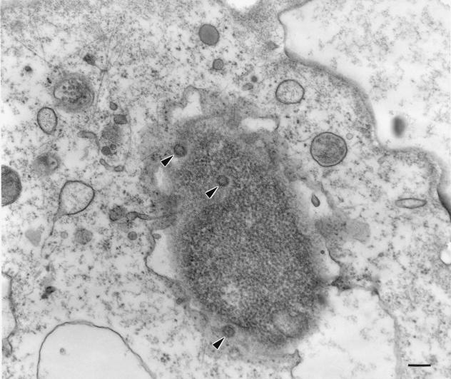
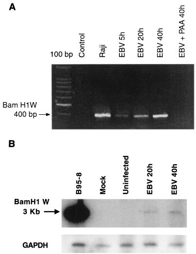
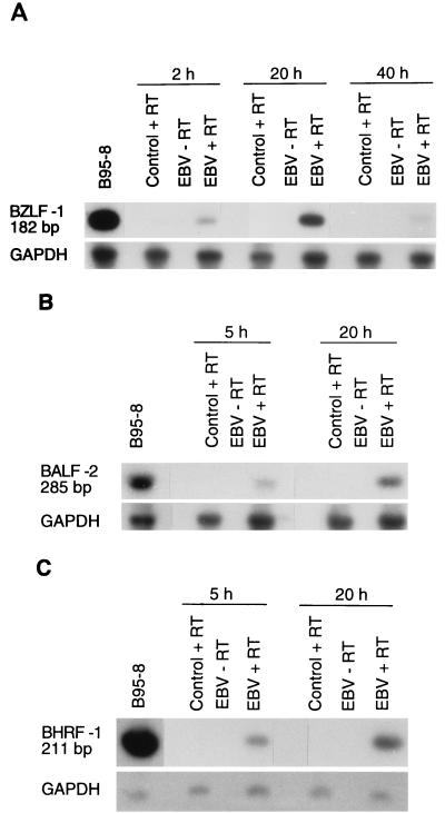
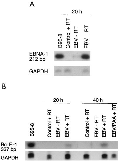
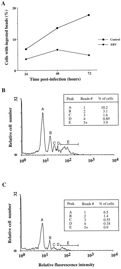
Previous studies have reported that infection of monocytes by viruses such as cytomegalovirus and human immunodeficiency virus weakens host natural immunity. In the present study, we demonstrated the capability of Epstein-Barr virus (EBV) to infect and replicate in freshly isolated human monocytes. Using electron microscopy analysis, we observed the presence of EBV virions in the cytoplasm and nuclei of approximately 20% of monocytes. This was confirmed by Southern blot analysis of EBV genomic DNA sequences in isolated nuclei from monocytes. Infection of monocytes by EBV leads to the activation of the replicative cycle. This was supported by the detection of immediate-early lytic mRNA BZLF-1 transcripts, and by the presence of two early lytic transcripts (BALF-2, which appears to function in DNA replication, and BHRF-1, also associated with the replicative cycle). The late lytic BcLF-1 transcripts, which code for the major nucleocapsid protein, were also detected, as well as EBNA-1 transcripts. However, attempts to detect EBNA-2 transcripts have yielded negative results. Viral replication was also confirmed by the release of newly synthesized infectious viral particles in supernatants of EBV-infected monocytes. EBV-infected monocytes were found to have significantly reduced phagocytic activity, as evaluated by the quantification of ingested carboxylated fluoresceinated latex beads. Taken together, our results suggest that EBV infection of monocytes and alteration of their biological functions might represent a new mechanism to disrupt the immune response and promote viral propagation during the early stages of infection.


Figures

FIG. 1
FIG. 1
EBV infection of human monocytes. Monocytes were incubated on ice in the presence or absence of infectious EBV for 5 min and then cultured at 37°C for 45 min. The preparation and examination of samples was performed as described in Materials and Methods. Virions are indicated by arrows (magnification of ×45,000). Black bar = 200 nm.
FIG. 2
FIG. 2
Detection of EBV genome in infected monocytes. Monocytes (107 cells) were treated with the phagocytosis inhibitor cytochalasin B (10 μM) for 10 min and infected with EBV for the indicated times. (A) The presence of EBV genome in purified cell nuclei was evaluated by PCR amplification of the BamHI W fragment, and the resulting 400-bp PCR product was visualized by ethidium bromide staining on a 2% agarose gel. In some samples, monocytes were also pretreated with PAA (200 μg/ml) before EBV infection. The Raji cell line was used as a positive control, and noninfected monocytes were used as negative controls. The first lane on the left represents a 100-bp DNA ladder. (B) Genomic DNA isolated from noninfected or EBV-infected monocytes (10 μg) or the Raji cell line (2 μg) was digested with BamHI and subjected to Southern blot analysis with a 32P-labelled BamHI-W probe. The 3-kb hybridization signal (BamHI-W fragment) obtained from the Raji cell line and monocytes previously infected with EBV for 20 and 40 h (EBV 20h, EBV 40h) are shown. Results are representative of three different experiments. YAC-1 cells were used as negative controls.
FIG. 3
FIG. 3
Detection of immediate-early and early transcripts by RT-PCR analysis. Total RNA was isolated from enriched monocytes (107 cells) exposed to EBV for 2 h and cultured for the indicated time periods. Following treatment with DNase I, RNA was reverse transcribed and amplified with sets of PCR primers specific for each gene (see Table 1). The size of the amplified fragments was 182 bp for BZLF-1 (A), 285 bp for BALF-2 (B), and 211 bp for BHRF-1 (C). PCR products were hybridized by Southern blot analysis using specific probes. GAPDH cDNA was used as an internal control. Tetradecanoyl phorbol acetate-treated B95-8 cells were used as positive controls, and noninfected monocytes were used as negative controls. The results presented are from one experiment and are representative of three separate experiments.
FIG. 4
FIG. 4
Detection of EBNA-1 and late lytic transcripts by RT-PCR analysis. Total RNA was isolated from enriched monocytes (107 cells), infected with EBV for 2 h, and cultured for the indicated periods of time. Following treatment with DNase I, RNA was reverse transcribed and amplified with sets of PCR primers specific for each gene. The size of the amplified fragments was 212 bp for EBNA-1 (A) and 332 bp for BcLF-1 (B). PCR products were hybridized with specific probes. GAPDH cDNA was used as an internal control. Tetradecanoyl phorbol acetate-treated B95-8 cells were used as positive controls, and noninfected monocytes were used as negative controls. Results are from one experiment and are representative of three separate experiments.
FIG. 5
FIG. 5
Suppression of phagocytosis in EBV-infected monocytes. EBV-infected or uninfected monocytes (5 × 10⁵ cells) were incubated with carboxylated fluoresceinated microspheres (at a ratio of 12 particles/cell), and the uptake of fluorescent particles was measured by flow cytometry, as described in Materials and Methods. (A) The percentage of fluorescence-positive cells (cells associated with at least one fluorescent microsphere) for EBV-infected and uninfected monocytes was measured at 24, 48, and 72 h postinfection. This time course analysis is representative of two experiments performed with two different healthy donors. Panels B and C are typical histograms showing the percentage of fluorescence-positive cells at each level of fluorescence intensity for uninfected and infected monocytes, respectively (this experiment was done at 60 h postinfection). Each peak is related to a definite number of fluorescent microspheres, and the percentage of positive cells contained in each peak is shown in the insert. A total of 104 cells was analyzed for each histogram. Results are representative of four other experiments.

References

    1. Adams D O, Hamilton T A. Macrophages as destructive cells in host defense. In: Gallin J I, Goldstein I M, Snyderman R, editors. Inflammation: basic principles in clinical correlates. 2nd ed. New York, N.Y: Raven Press; 1992. pp. 637–662.
    1. Andoh A, Fujiyama Y, Kitoh K, Hodohara K, Bamba T, Hosoda S. Flow cytometric assay for phagocytosis of human monocytes mediated via Fcγ-receptors and complement receptor CR1 (CD35) Cytometry. 1991;12:677–686. - PubMed
    1. Baer R, Bankier A T, Biggin M D, Deininger P L, Farrell P J, Gibson T J, Hatfull G, Hudson G S, Satchwell S C, Séguin C, Tuffnell P S, Barrell B G. DNA sequence and expression of the B95-8 Epstein-Barr virus genome. Nature. 1984;310:207–211. - PubMed
    1. Baldwin G C, Fleischmann J, Chung Y, Koyanagi Y, Chen I S, Golde D W. Human immunodeficiency virus causes mononuclear phagocyte dysfunction. Proc Natl Acad Sci USA. 1990;87:3933–3937. - PMC - PubMed
    1. Beaulieu A D, Paquin R, Gosselin J. Epstein-Barr virus modulates de novo protein synthesis in human neutrophils. Blood. 1995;86:2789–2798. - PubMed

Publication types

Substances

LinkOut - more resources