. 2000 Mar;74(6):2721-30.
doi: 10.1128/jvi.74.6.2721-2730.2000.

Activation of lymphocyte signaling by the R1 protein of rhesus monkey rhadinovirus

Affiliations

Activation of lymphocyte signaling by the R1 protein of rhesus monkey rhadinovirus

B Damania et al. J Virol. 2000 Mar.

Abstract

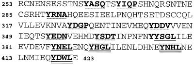
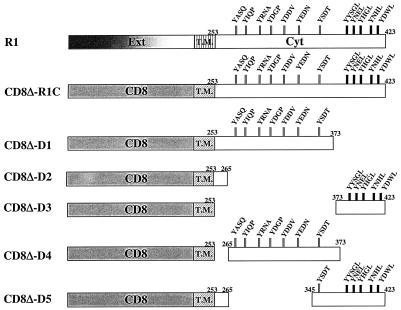
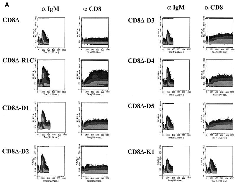
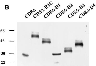
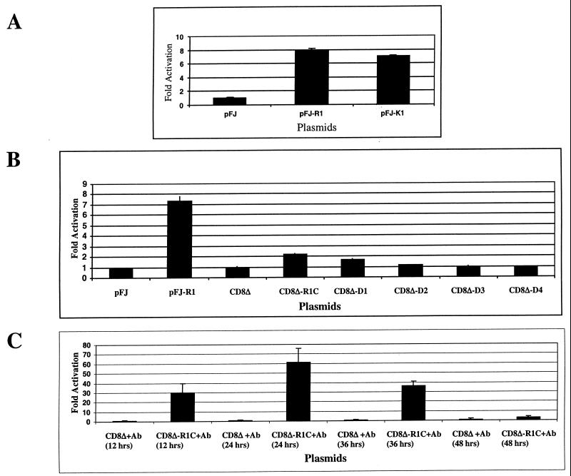
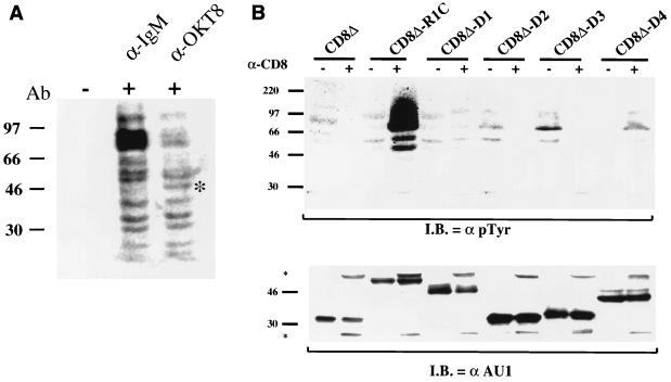
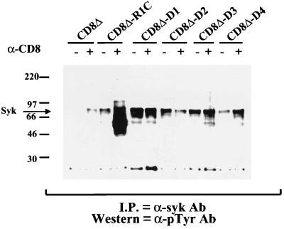
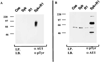
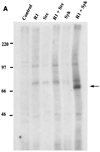
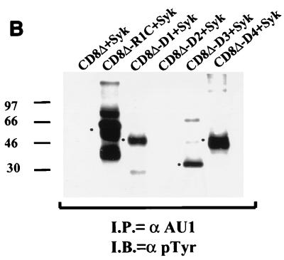
Rhesus monkey rhadinovirus (RRV) is a gamma-2 herpesvirus that exhibits a considerable degree of similarity to the human Kaposi's sarcoma-associated herpesvirus (KSHV). The R1 protein of RRV is distantly related to the K1 protein of KSHV, and R1, like K1, can contribute to cell growth transformation. In this study we analyzed the ability of the cytoplasmic tail of R1 to function as a signal transducer. The cytoplasmic domain of the R1 protein contains several tyrosine residues whose phosphorylation is induced in cells expressing Syk kinase. Expression of a CD8 chimera protein containing the extracellular and transmembrane domains of CD8 fused to the cytoplasmic domain of R1 mobilized intracellular calcium and induced cellular tyrosine phosphorylation in B cells upon stimulation with anti-CD8 antibody. None of the CD8-R1 cytoplasmic deletion mutants tested were able to mobilize intracellular calcium or to induce tyrosine phosphorylation to a significant extent upon addition of anti-CD8 antibody. Expression of wild-type R1 protein activated nuclear factor of activated T lymphocytes (NFAT) eightfold in B cells in the absence of antibody stimulation; expression of the CD8-R1C chimera strongly induced NFAT activity (60-fold) but only upon the addition of anti-CD8 antibody. We conclude that the cytoplasmic domain of R1 is capable of transducing signals that elicit B-lymphocyte activation events. The signal-inducing properties of R1 appear to be similar to those of K1 but differ in that the required sequences are distributed over a much longer stretch of the cytoplasmic domain (>150 amino acids). In addition, the induction of calcium mobilization was considerably longer in duration and stronger with R1 than with K1.


Figures

FIG. 1
FIG. 1
Amino acid sequences of the R1 cytoplasmic tail. The 170 amino acids of the C-terminal cytoplasmic domain of R1 are shown. The cytoplasmic domain of R1 contains several potential SH2 binding motifs (boldface). Dotted underlining, SH2 binding motifs YXXA, YXXV, YXXT, and YXXP; solid underlining, last five YXXL motifs. The third and fourth and fourth and fifth YXXL motifs resemble ITAMs in that they and the surrounding sequences are spaced in a fashion consistent with the consensus sequence, (D/E)X7(D/E)X2YX2LX7–10YX2L/I. These motifs are boxed.
FIG. 2
FIG. 2
Construction of CD8-R1 chimeras. The cytoplasmic domain (Cyt) of R1 was fused to the extracellular (Ext) and transmembrane (T.M.) domains of CD8. The CD8 chimera containing the full-length cytoplasmic domain of R1 (CD8Δ-R1C) and cytoplasmic domains with various deletions (CD8Δ-D1, CD8Δ-D2, CD8Δ-D3, CD8Δ-D4, CD8Δ-D5) are depicted. YXXX, tyrosine residues and their surrounding sequences.
FIG. 3
FIG. 3
Mobilization of intracellular free calcium upon antibody stimulation. (A) Intracellular calcium release from the chimera cell lines was monitored by flow cytometry. Either an anti-IgM antibody (left) or an anti-CD8 antibody (right) was used to stimulate cells. Data are presented as histograms of the numbers of cells expressing blue fluorescence (y axis) versus time. The intensities of the responses are depicted as different shades of gray, with the darkest shade representing the highest levels of free calcium in the cell. (B) Western blot analysis of CD8-R1 chimera cell extracts using an anti-AU1 antibody to detect expression levels of the AU1-tagged chimera proteins.
FIG. 3
FIG. 3
Mobilization of intracellular free calcium upon antibody stimulation. (A) Intracellular calcium release from the chimera cell lines was monitored by flow cytometry. Either an anti-IgM antibody (left) or an anti-CD8 antibody (right) was used to stimulate cells. Data are presented as histograms of the numbers of cells expressing blue fluorescence (y axis) versus time. The intensities of the responses are depicted as different shades of gray, with the darkest shade representing the highest levels of free calcium in the cell. (B) Western blot analysis of CD8-R1 chimera cell extracts using an anti-AU1 antibody to detect expression levels of the AU1-tagged chimera proteins.
FIG. 4
FIG. 4
Activation of NFAT activity in BJAB cells by the R1 protein. (A) BJAB cells were transfected with an NFAT luciferase and β-galactosidase reporter plasmid along with either a pFJ vector control plasmid, a pFJ-R1-expressing plasmid, or a pFJ-K1-expressing plasmid. At 48 h posttransfection cells were harvested and tested for luciferase activity. Luciferase counts for each sample were normalized with respect to β-galactosidase activity. Luciferase assays were repeated three times, and the average of these values was graphed as the fold activation of NFAT by the R1 and K1 proteins versus that of control vector alone. (B) Transfections similar to those in panel A except that the CD8Δ-R1 chimera-expressing plasmids indicated in the graph were electroporated into BJAB cells. (C) BJAB cells were transfected with NFAT luciferase and β-galactosidase reporter plasmids along with a CD8Δ-R1C-expressing plasmid. At 24 h posttransfection cells were harvested and incubated with anti-CD8 antibody for 12, 24, 36, and 48 h. Ab, antibody.
FIG. 5
FIG. 5
Induction of cellular tyrosine phosphorylation upon antibody stimulation. (A) Comparison of anti-IgM- and anti-CD8-stimulated CD8Δ-R1C cells. Cross-linking of the CD8Δ-R1C cell line with an anti-IgM or anti-CD8 (OKT8) antibody (Ab) induced tyrosine phosphorylation of cellular proteins as determined by Western analysis using an anti-pTyr antibody. Asterisk, 50-kDa phosphorylated protein seen in the CD8 antibody-stimulated, but not IgM antibody-stimulated, B cells. (B) Mutational analysis of CD8Δ-R1 chimeras for the induction of tyrosine phosphorylation upon antibody stimulation. Cells were incubated in the absence (−) or presence (+) of anti-CD8 antibody for 1 min at 37°C and immediately lysed. Cell extracts were subjected to gel electrophoresis, transferred to nitrocellulose, and reacted with an anti-pTyr antibody (top). Cell extracts were probed with an anti-AU1 antibody (bottom) to show comparative levels of chimeric proteins in these cells. Asterisks, heavy and light chains of the CD8 antibody used for stimulation. I.B., immunoblotting.
FIG. 6
FIG. 6
Tyrosine phosphorylation of Syk kinase and CD8 chimeras. Shown is the induction of tyrosine phosphorylation by Syk kinase. The CD8-R1 chimera cell lines were incubated with (+) or without (−) anti-CD8 antibody. Extracts were subjected to immunoprecipitation (I.P.) with an anti-Syk antibody (Ab), and immune complexes were resolved by gel electrophoresis and subjected to a Western blot analysis using an anti-pTyr antibody.
FIG. 7
FIG. 7
Syk kinase induces the phosphorylation of full-length R1. (A) Cotransfection of R1 and Syk expression plasmids in Cos-1 cells. A full-length, C-terminal AU1 epitope-tagged R1-expressing plasmid was transfected into Cos-1 cells either alone or in the presence of Syk kinase. Cells were harvested 48 h posttransfection, and extracts were subjected to immunoprecipitation (I.P.) with an anti-AU1 antibody followed by a Western blot analysis (I.B.) using an anti-pTyr antibody. (B) The same extracts were coimmunoprecipitated with an anti-pTyr antibody followed by Western blotting with an anti-AU1 antibody.
FIG. 8
FIG. 8
Syk-induced phosphorylation of R1 and CD8Δ-R1 chimeras. (A) Phosphorylation of R1 in vitro. An in vitro kinase assay was performed using extracts from Cos-1 cells transfected with R1 alone or in combination with Syk and Src kinase. Immune complexes were washed extensively and subsequently incubated with [γ-32P]ATP. Complexes were then separated by gel electrophoresis and exposed to autoradiography. Arrow, presence of a 70 kDa phosphorylated protein that corresponds to the molecular sizes of both Syk and R1. (B) Tyrosine phosphorylation of CD8Δ-R1 chimeras induced by Syk kinase. Cos-1 cells were cotransfected with a Syk expression plasmid along with the CD8Δ-R1 chimera-expressing plasmids. The chimeras were tagged with an anti-AU1 antibody. Immunoprecipitations (I.P.) were performed using an anti-AU1 antibody and an anti-pTyr antibody was used for a Western blot analysis (I.B.). Dots, positions of mutant proteins.
FIG. 8
FIG. 8
Syk-induced phosphorylation of R1 and CD8Δ-R1 chimeras. (A) Phosphorylation of R1 in vitro. An in vitro kinase assay was performed using extracts from Cos-1 cells transfected with R1 alone or in combination with Syk and Src kinase. Immune complexes were washed extensively and subsequently incubated with [γ-32P]ATP. Complexes were then separated by gel electrophoresis and exposed to autoradiography. Arrow, presence of a 70 kDa phosphorylated protein that corresponds to the molecular sizes of both Syk and R1. (B) Tyrosine phosphorylation of CD8Δ-R1 chimeras induced by Syk kinase. Cos-1 cells were cotransfected with a Syk expression plasmid along with the CD8Δ-R1 chimera-expressing plasmids. The chimeras were tagged with an anti-AU1 antibody. Immunoprecipitations (I.P.) were performed using an anti-AU1 antibody and an anti-pTyr antibody was used for a Western blot analysis (I.B.). Dots, positions of mutant proteins.
FIG. 9
FIG. 9
R1 can specifically interact with Syk kinase. Coimmunoprecipitations (I.P.) were performed using an anti-Src or anti-Syk kinase antibody on cells transfected with an AU1-tagged R1-expressing plasmid alone or together with Src or Syk kinase-expressing plasmids. Immune complexes were subjected to gel electrophoresis. The gel was transferred to nitrocellulose and subsequently probed with an anti-AU1 antibody. Bottom, levels of Syk and Src kinase in transfected cells. I.B., immunoblotting.
FIG. 10
FIG. 10
Tyrosine phosphorylation of the R1 and K1 proteins. Comparison of the phosphorylation of R1 and K1 proteins by Src and Syk kinase. Both R1- and K1-expressing plasmids were transfected into Cos-1 cells in the absence or presence of Src and Syk kinases. The immunoblot (I.B.) was reacted with a pTyr antibody (Top). Arrows, R1 and K1 proteins. (Bottom) Western blot analysis of cell extracts used for the immunoprecipitations in the upper panel. The extracts were probed with an AU1 antibody to detect expression levels of R1 and K1 proteins expressed in transfected cells.

References

    1. Alexander, L., L. Denenkamp, A. Knapp, M. Auerbach, S. Czajak, B. Damania, and R. C. Desrosiers. 1999. The primary sequence of rhesus rhadinovirus isolate 26-95: sequence similarities to Kaposi's sarcoma herpesvirus and rhesus rhadinovirus isolate 17577. J. Virol. - PMC - PubMed
    1. Beaufils P, Choquet D, Mamoun R Z, Malissen B. The (YXXL/I)2 signaling motif found in the cytoplasmic segments of the bovine leukaemia virus envelope protein and Epstein-Barr virus latent membrane protein 2A can elicit early and late lymphocyte activation events. EMBO J. 1993;12:5105–5112. - PMC - PubMed
    1. Cambier J C. Antigen and Fc receptor signaling. The awesome power of the immunoreceptor tyrosine-based activation motif (ITAM) J Immunol. 1995;155:3281–3285. - PubMed
    1. Cambier J C, Jensen W A. The hetero-oligomeric antigen receptor complex and its coupling to cytoplasmic effectors. Curr Opin Genet Dev. 1994;4:55–63. - PubMed
    1. Cambier J C, Pleiman C M, Clark M R. Signal transduction by the B cell antigen receptor and its coreceptors. Annu Rev Immunol. 1994;12:457–486. - PubMed

Publication types

MeSH terms