. 2000 Mar;74(6):2760-9.
doi: 10.1128/jvi.74.6.2760-2769.2000.

Functional characterization of the human immunodeficiency virus type 1 genome by genetic footprinting

Affiliations

Functional characterization of the human immunodeficiency virus type 1 genome by genetic footprinting

L C Laurent et al. J Virol. 2000 Mar.

Abstract

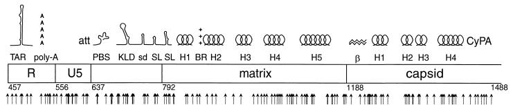
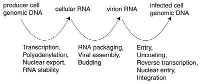
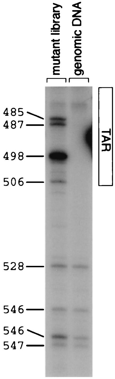
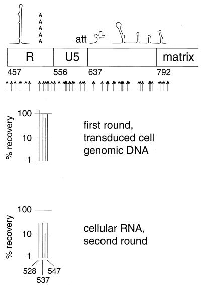
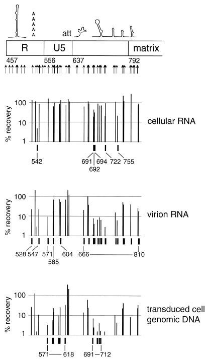
We present a detailed and quantitative analysis of the functional characteristics of the 1,000-nucleotide segment at the 5' end of the human immunodeficiency virus type 1 (HIV-1) RNA genome. This segment of the viral genome contains several important cis-acting sequences, including the TAR, polyadenylation, viral att site, minus-strand primer-binding site, and 5' splice donor sequences, as well as coding sequences for the matrix protein and the N-terminal half of the capsid protein. The genetic footprinting technique was used to determine quantitatively the abilities of 134 independent insertion mutations to (i) make stable viral RNA, (ii) assemble and release viral RNA-containing viral particles, and (iii) enter host cells, complete reverse transcription, enter the nuclei of host cells, and generate proviruses in the host genome by integration. All of the mutants were constructed and analyzed en masse, greatly decreasing the labor typically involved in mutagenesis studies. The results confirmed the presence of several previously known functional features in this region of the HIV genome and provided evidence for several novel features, including newly identified cis-acting sequences that appeared to contribute to (i) the formation of stable viral transcripts, (ii) viral RNA packaging, and (iii) an early step in viral replication. The results also pointed to an unanticipated trans-acting role for the N-terminal portion of matrix in the formation of stable viral RNA transcripts. Finally, in contrast to previous reports, the results of this study suggested that detrimental mutations in the matrix and capsid proteins principally interfered with viral assembly.


Figures

FIG. 1
FIG. 1
Mutagenesis scheme using MuA tranposase. Oligonucleotides used for mutagenesis are represented by bold lines (formula image
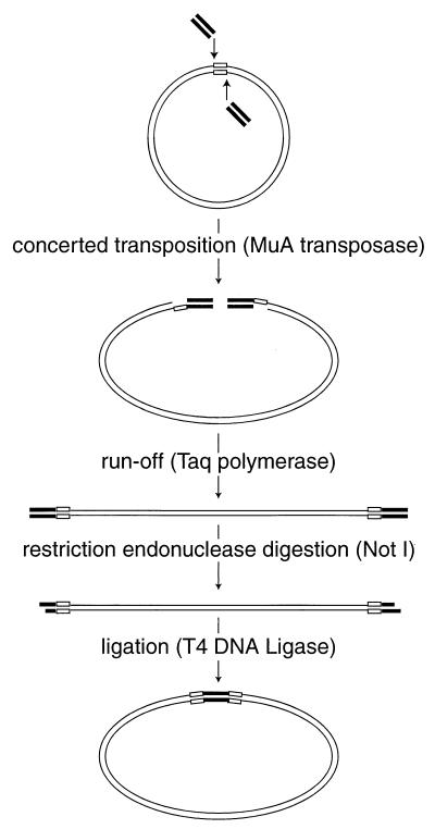
), the target DNA plasmid is represented by thin lines, and the 5-bp sequences in the target DNA that were duplicated during mutagenesis are represented by open boxes (formula image).
FIG. 2
FIG. 2
Map of the mutations evaluated in this study. Previously described features, indicated on the map, are TAR, the polyadenylation signal (poly-A), the att site (att), the primer-binding site (PBS), the kissing loop domain (KLD), the major splice donor (sd), two Gag-binding stem-loop structures (SL), alpha helices 1 to 5 (H1 to H5) of matrix, the basic region of matrix (BR), the beta hairpin of capsid (β), helices 1 to 4 (H1 to H4) of capsid, and the cyclophilin A-binding region of capsid (CyPA). Numbers indicate nucleotide positions at the borders of major regions in the HIV genome. Arrows indicate positions of insertional mutations evaluated in this study.
FIG. 3
FIG. 3
Relationship of nucleic acid samples analyzed by genetic footprinting to specific steps in the life cycle. A decrease in the abundance of a given mutant between two of the indicated nucleic acid samples implies that the mutant is defective in one of the intervening steps of the viral life cycle.
FIG. 4
FIG. 4
Design of transfection and transduction experiments. The library of mutagenized proviruses was either transiently or stably transfected into cells to produce populations of mutant virions. In our experiments, the viral genomes of mutants defective in trans-acting factors were efficiently rescued during the transient transfection by phenotypic mixing but were not detectably rescued during the stable transfection. The virus produced in the transient transfection experiment was used to transduce fresh, untransduced cells at a low multiplicity, resulting in a population of producer cells that contained a single provirus per cell. Symbols represent wild-type viral genome (formula image
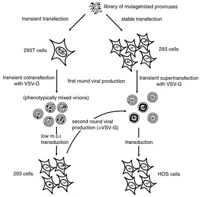
), replication-defective viral genome with mutation in trans-acting factor (formula image), wild-type viral protein (formula image), and mutant viral protein (formula image).
FIG. 5
FIG. 5
Genetic footprinting scheme using flanking PCR and restriction digestion. A collection of insertion mutants is subjected to PCR using one radioactively labeled primer (formula image
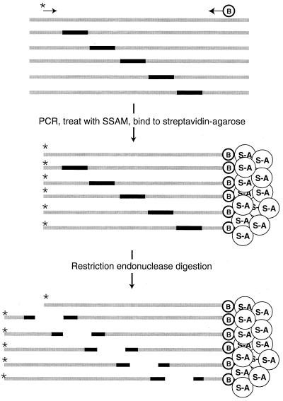
) and one biotinylated primer (formula image). The PCR products are captured by streptavidin-agarose resin (formula image) and digested with a restriction enzyme that recognizes a site in the insertion sequence. The radioactively labeled ends of the PCR products are released.
FIG. 6
FIG. 6
Genetic footprinting of library of mutagenized proviruses and nucleic acid samples from the transient transfection experiment, second round (the uncomplemented round) of viral production and transduction (cellular RNA, viral RNA, and transduced cell genomic DNA). Numbers directly to the left of the gel indicate exact positions of insertions. In this numbering convention, the first nucleotide of the HIV provirus is at position 1. Quantitative values for recovery of the mutants represented by each band, in each sample, averaged from normalized measurements are also given to the left of the gel. The nucleic acid sequences of the mutants represented by each band are written to the right of the gel. The sequences derived from the insertion oligonucleotide are boxed, and the target sequence duplications are underlined. Eighty-nine percent of 19 individually sequenced mutants contained the expected precise 5-bp duplication flanking the 9-bp insertion.
FIG. 7
FIG. 7
Percent recovery of mutants through single-cycle transductions. Data are not shown for mutants for which the coefficient of variation between triplicate experiments was greater than 0.5, except in cases where the observed phenotypes were confirmed by reanalysis. Ordinate values are plotted on a logarithmic scale. Tick marks indicate mutants that display severe depletions (<45% recovery). Mutations that compromise replication in both the presence and the absence of complementation are considered to be located in cis-acting sequences, while those that affect only uncomplemented replication cycles are considered to be located in trans-acting sequences. (A) Percent recovery of mutants after a single round of transduction in the presence of complementation. Data are from the transient transfection experiment, first round of transduction. (B) Percent recovery of mutants after a single round of transduction in the absence of complementation. Data are from the transient transfection experiment, second round of transduction. Data are not given for mutants whose abundance after the first round of infection was too low to allow accurate quantitation of further depletion.
FIG. 8
FIG. 8
Genetic footprinting of library of mutagenized proviruses and infected cell genomic DNA sample from the transient transfection experiment, first round (the complemented round) of viral production and transduction. Numbers directly to the left of the gel indicate exact positions of insertions. In this numbering convention, the first nucleotide of the HIV provirus is at position 1. The extent of the TAR region is indicated to the right of the gel.
FIG. 9
FIG. 9
Effects of selected mutations in cis-acting sequences in the transient transfection experiment. These mutants were replication competent in the first round of transduction but defective for transcript formation in the second round, possibly indicating that it is the copy of these cis-acting sequences in the 3′ LTR which is active.
FIG. 10
FIG. 10
Recovery of genomes carrying mutations in cis-acting sequences, at several steps of a single-cycle uncomplemented transduction. Samples were collected from the stable transfection experiment. Percent recovery was calculated by dividing the abundance of a mutant in a given nucleic acid sample by the abundance of that mutant in the genomic DNA sample from the stable transfection. Data are not shown for points where the abundance of a particular mutant was very low in the preceding nucleic acid sample or where the coefficient of variation between triplicate experiments was greater than 0.5. Graphs are plotted on a log scale. Tick marks indicate mutants that display severe depletions (<50% of preceding nucleic acid sample). Mutants which were depleted in the cellular RNA sample are considered to be defective in transcript formation, mutants which were depleted in the virion RNA sample are considered to be defective in packaging, and mutants which were depleted in the transduced cell genomic DNA sample are considered to be defective in an early step in viral transduction.
FIG. 11
FIG. 11
Recovery of mutations in matrix and capsid at several steps of a single-cycle uncomplemented transduction. Samples were collected from the transient transfection experiment, second round of viral production and transduction. Percent recovery was calculated by dividing the abundance of a mutant in a given nucleic acid sample by the abundance of that mutant in the transduced cell genomic DNA sample from the first round of transduction. Data are not shown for points where the abundance of a particular mutant was very low in the preceding nucleic acid sample or where the coefficient of variation between triplicate experiments was greater than 0.5. Graphs are plotted on a log scale. Tick marks indicate mutants that display severe depletions (<45% of preceding nucleic acid sample). Mutants which were depleted in the cellular RNA sample are considered to be defective in transcript formation, mutants which were depleted in the virion RNA sample are considered to be defective in assembly, and mutants which were depleted in the transduced cell genomic DNA sample are considered to be defective in an early step in viral replication. All mutations in capsid that affected viral replication showed their effects in trans.
FIG. 12
FIG. 12
Percent recovery of matrix and capsid mutants through the viral assembly process and the early steps of the viral life cycle. Data are derived from single-round uncomplemented viral production and transduction cycles. A schematic of the phases of the life cycle tested is drawn above the graphs. Numbers to the right of each point indicate the number of mutants from which data were averaged. Below each data point is a schematic of the region of matrix or capsid evaluated. Error bars indicate 95% confidence intervals. (A) Data for the matrix protein. Data are shown for insertions in the complete matrix protein (formula image 
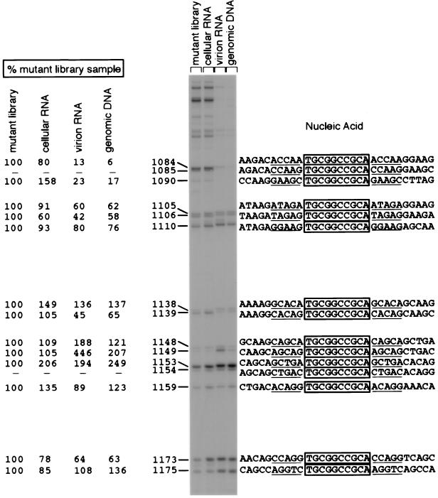
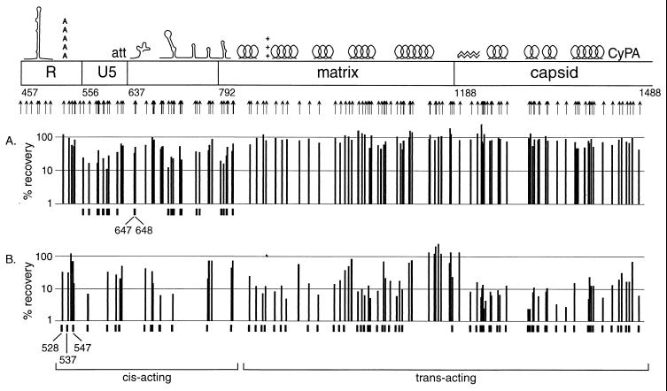
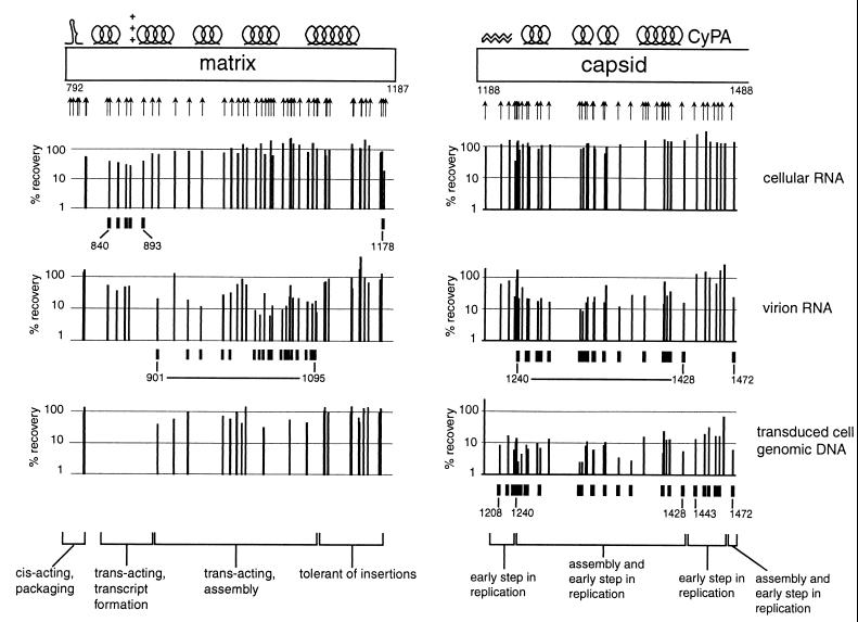
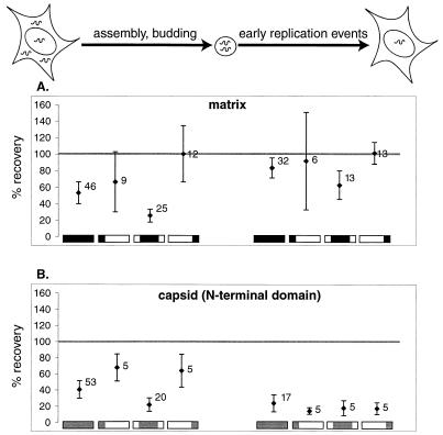
[amino acids 1 to 132; nucleotides 792 to 1187]), the N-terminal region (formula image [amino acids 1 to 35; nucleotides 792 to 896]), the central region (formula image [amino acids 36 to 102; nucleotides 897 to 1097]), and the C-terminal region (formula image [amino acids 104 to 130; nucleotides 1101 to 1181]). (B) Data for the N-terminal half of the capsid protein. Data are shown for insertions in the N-terminal half (formula image [amino acids 1 to 101; nucleotides 1188 to 1490]), the N-terminal beta hairpin (formula image [amino acids 1 to 15; nucleotides 1188 to 1232]), the central helical region (formula image [amino acids 17 to 81, nucleotides 1236 to 1430]), and the cyclophilin A-binding region (formula image [amino acids 85 to 96; nucleotides 1440 to 1475]).

References

    1. Baker T A, Mizuuchi M, Savilahti H, Mizuuchi K. Division of labor among monomers within the Mu transposase tetramer. Cell. 1993;74:723–733. - PubMed
    1. Berkowitz R D, Luban J, Goff S P. Specific binding of human immunodeficiency virus type 1 Gag polyprotein and nucleocapsid protein to viral RNAs detected by RNA mobility shift assays. J Virol. 1993;67:7190–7200. - PMC - PubMed
    1. Berkowitz R D, Goff S P. Analysis of binding elements in the human immunodeficiency virus type 1 genomic RNA and nucleocapsid protein. Virology. 1994;202:233–246. - PubMed
    1. Braaten D, Franke E K, Luban J. Cyclophilin A is required for an early step in the life cycle of human immunodeficiency virus type 1 before the initiation of reverse transcription. J Virol. 1996;70:3551–3560. - PMC - PubMed
    1. Bukrinskaya A G, Vorkunova G K, Tentsov Y. HIV-1 matrix protein p17 resides in cell nuclei in association with genomic RNA. AIDS Res Hum Retroviruses. 1992;8:1795–1801. - PubMed

Publication types