. 2000 Mar;74(6):2777-85.
doi: 10.1128/jvi.74.6.2777-2785.2000.

Infectious entry pathway of adeno-associated virus and adeno-associated virus vectors

Affiliations

Infectious entry pathway of adeno-associated virus and adeno-associated virus vectors

J S Bartlett et al. J Virol. 2000 Mar.

Abstract

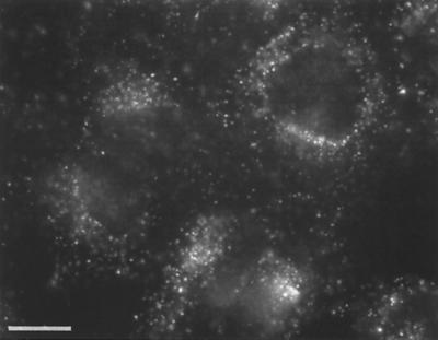
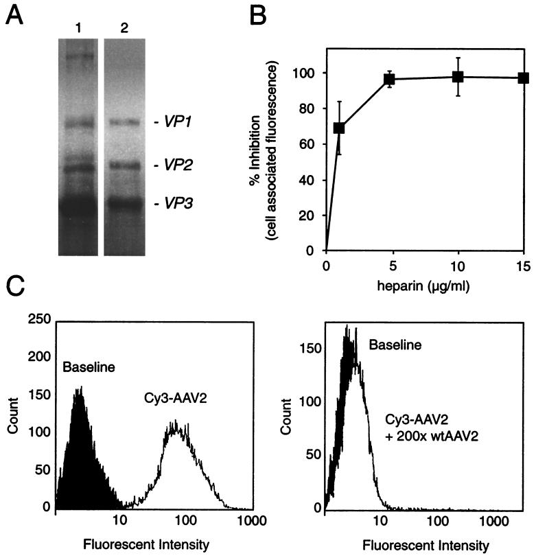
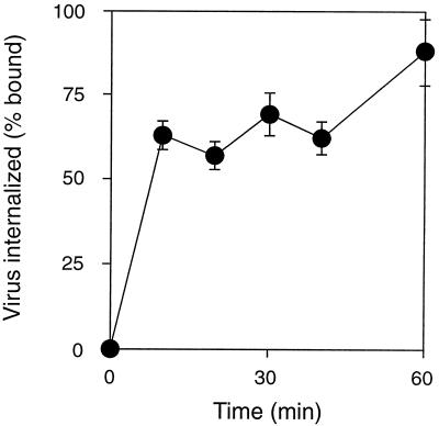
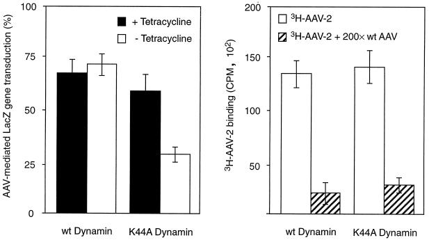
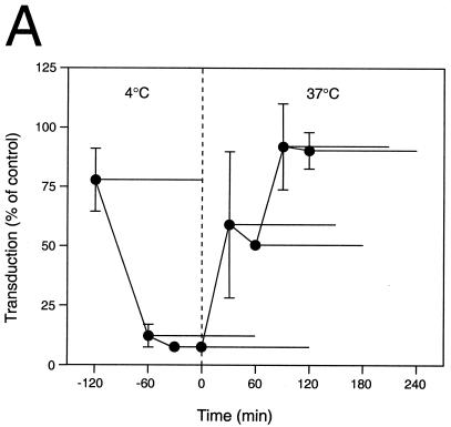
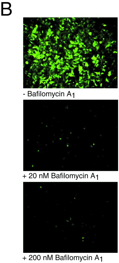
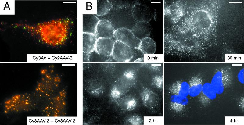
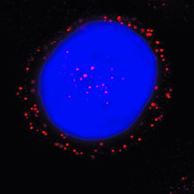
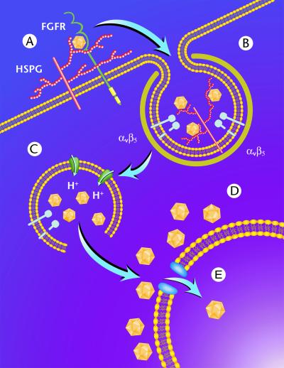
We have investigated the infectious entry pathway of adeno-associated virus (AAV) and recombinant AAV vectors by assessing AAV-mediated gene transfer and by covalently conjugating fluorophores to AAV and monitoring entry by fluorescence microscopy. We examined AAV entry in HeLa cells and in HeLa cell lines which inducibly expressed a dominant interfering mutant of dynamin. The data demonstrate that AAV internalizes rapidly by standard receptor-mediated endocytosis from clathrin-coated pits (half-time <10 min). The lysosomotropic agents ammonium chloride and bafilomycin A(1) prevent AAV-mediated gene transfer when present during the first 30 min after the onset of endocytosis, indicating that AAV escapes from early endosomes yet requires an acidic environment for penetration into the cytosol. Following release from the endosome, AAV rapidly moves to the cell nucleus and accumulates perinuclearly beginning within 30 min after the onset of endocytosis. We present data indicating that escape of AAV from the endosome and trafficking of viral particles to the nucleus are unaffected by the presence of adenovirus, the primary helper virus for a productive AAV infection. Within 2 h, viral particles could be detected within the cell nucleus, suggesting that AAV enters the nucleus prior to uncoating. Interestingly, the majority of the intracellular virus particles remain in a stable perinuclear compartment even though gene expression from nuclear AAV genomes can be detected. This suggests that the process of nuclear entry is rate limiting or that AAV entry involves multiple pathways. Nevertheless, these data establish specific points in the AAV infectious entry process and have allowed the generation of a model for future expansion to specific cell types and AAV vector analysis in vivo.


Figures

FIG. 1
FIG. 1
Uptake of Cy3AAV-2 by HeLa cells. HeLa cells were incubated with 1.25 × 10¹¹ particles of Cy3AAV-2/ml for 10 min at 37°C, washed to remove virus particles that had not internalized, and maintained at 37°C for 30 min. Labeled virus was visualized by fluorescence microscopy. Bar, 10 μm.
FIG. 2
FIG. 2
Characterization of Cy3-labeled AAV particles: Cy3AAV-2 maintains dependence on HSPG-mediated attachment. (A) SDS-PAGE analysis of labeled capsid proteins (lane 1) and unmodified capsid proteins (lane 2). Protein cross-linking due to the labeling reaction was less than 5%. (B) Quantitative assessment of cell-associated Cy3 fluorescence intensity following coincubation of HeLa cells with Cy3AAV-2 and increasing amounts of heparin. (C) Assessment of cell-associated Cy3 fluorescence by fluorescence-activated cell sorter analysis. HeLa cells were incubated with 2.5 × 10¹⁰ particles of Cy3AAV-2/ml for 30 min at 4°C (left panel) or with 2.5 × 10¹⁰ particles of Cy3AAV-2/ml in the presence of 5 × 10¹² particles of unlabeled AAV-2 (wtAAV2) for 30 min at 4°C (right panel). Both sets of cells were then washed, fixed, and directly subjected to flow-cytometric analysis.
FIG. 3
FIG. 3
Internalization of AAV-2 by HeLa cells. HeLa cells were incubated with [3H]AAV-2 at 4°C for 60 min, washed to remove unattached virus, and then incubated at 37°C for various lengths of time (0 to 90 min). Virus that had not internalized was removed from the cell surface by washing with a mildly acidic buffer, and cell-associated radioactivity was determined by scintillation counting. Values shown are means ± standard error and have been adjusted by subtraction of background radioactivity (n = 3).
FIG. 4
FIG. 4
Entry of AAV-2 is mediated by dynamin-associated uptake pathways. AAV-2-mediated gene transfer (left panel) and AAV-2 attachment (right panel) to HeLa cells overexpressing wild-type (wt) dynamin were compared to those of cells expressing mutant (K44A) dynamin. Cells were exposed to [3H]AAV-2 (1010 particles/ml) or AAVlacZ (108 particles/ml) for 2 h at 4°C, and either attachment (3HAAV-2) was measured immediately or gene transfer (AAVlacZ) was assessed 48 h later. Expression of wild-type or mutant dynamin was induced by removal of tetracycline from the growth medium, as described in Materials and Methods. (Left panel) AAV-2-mediated gene transfer is shown in the presence and absence of tetracycline; (right panel). AAV-2 binding is shown in the absence of tetracycline. The specificity of AAV-2 binding was determined by competition with a 200-fold molar excess of unlabeled virus (wt AAV). Values shown are means ± standard error (n = 6 for each).
FIG. 5
FIG. 5
Inhibitory effect of ammonium chloride and bafilomycin A1 on AAV-mediated gene transfer. (A) AAVlacZ vector was bound to HeLa cells at 4°C for 120 min, and unbound virions were washed away. Ammonium chloride (25 mM final concentration) was added at the indicated time points (●) and was present during different 2-h periods as indicated by the horizontal bars. The ammonium chloride was washed out of the cells at the ends of these periods, and gene transfer was assessed at 24 h postinfection by X-Gal histochemistry. Values shown are means ± standard error (n = 3). (B) HeLa cells grown on chambered slides were preincubated without (−) or with bafilomycin A1 (20 or 200 nM) for 30 min at 37°C. AAVEGFP vector (1010 particles/ml) was allowed to bind to the cells for 10 min at 37°C, the cells were washed to remove unbound virus, and vector-mediated green fluorescent protein fluorescence was assessed by fluorescence microscopy 24 h later. Where indicated, bafilomycin A1 was present throughout the experiment. High-level AAV-mediated gene expression was restricted to HeLa cells grown in the absence of bafilomycin A1.
FIG. 5
FIG. 5
Inhibitory effect of ammonium chloride and bafilomycin A1 on AAV-mediated gene transfer. (A) AAVlacZ vector was bound to HeLa cells at 4°C for 120 min, and unbound virions were washed away. Ammonium chloride (25 mM final concentration) was added at the indicated time points (●) and was present during different 2-h periods as indicated by the horizontal bars. The ammonium chloride was washed out of the cells at the ends of these periods, and gene transfer was assessed at 24 h postinfection by X-Gal histochemistry. Values shown are means ± standard error (n = 3). (B) HeLa cells grown on chambered slides were preincubated without (−) or with bafilomycin A1 (20 or 200 nM) for 30 min at 37°C. AAVEGFP vector (1010 particles/ml) was allowed to bind to the cells for 10 min at 37°C, the cells were washed to remove unbound virus, and vector-mediated green fluorescent protein fluorescence was assessed by fluorescence microscopy 24 h later. Where indicated, bafilomycin A1 was present throughout the experiment. High-level AAV-mediated gene expression was restricted to HeLa cells grown in the absence of bafilomycin A1.
FIG. 6
FIG. 6
Pulse-labeling evaluation of fluorescent AAV distribution in HeLa cells, demonstrating the lack of intraendosomal colocalization of AAV and adenovirus following endocytosis and time-dependent translocation of AAV from the cell membrane to the perinuclear region. (A) HeLa cells were incubated for 10 min at 37°C with 1.25 × 10¹¹ particles of Cy2AAV-3/ml plus either 1.25 × 10¹¹ particles of Cy3Ad (top panel) or 1.25 × 10¹¹ particles of Cy3AAV-2 (lower panel)/ml and assessed for colocalization by fluorescence microscopy. Colocalization of serotype 2 and 3 AAV, but not of adenovirus and AAV, was evidenced by yellow signal from overlapping red (Cy3) and green (Cy2) signals. Bar, 10 μm. (B) HeLa cells were incubated with 1.25 × 10¹¹ particles of Cy3AAV-2/ml for 2 h at 4°C, washed, then incubated at 37°C for 0, 30, 120, or 240 min and assessed by fluorescence microscopy. The positions of nuclei are evident in the 4-h panel due to DAPI (blue) staining. Bar, 10 μm.
FIG. 7
FIG. 7
Distribution of Cy3AAV-2 particles in HeLa cells 2 h postinfection. HeLa S3 cells were pulsed-labeled with 1.25 × 10¹¹ particles of Cy3AAV-2 (red)/ml for 10 min at 37°C, washed to remove uninternalized virus, and incubated at 37°C for 2 h prior to analysis by confocal microscopy. The position of the cell nucleus was assessed by DAPI (blue) staining. A representative image is shown, consisting of a single plane of focus through the center of a cell.
FIG. 8
FIG. 8
Schematic representation of AAV entry and endocytic trafficking in HeLa cells. Following binding to cell surface HSPG (A), AAV is rapidly internalized via clathrin-coated pits (B) through a process involving αvβ5 integrin. Once internalized, the virus encounters a weakly acidic environment which is sufficient to allow penetration into the cytosol (C). Following endosome release, AAV accumulates perinuclearly (D) and slowly penetrates through the NPC into the nucleus (E).

Similar articles

Cited by

References

    1. Bartlett J S, Samulski R J. Fluorescent viral vectors: a new technique for the pharmacological analysis of gene therapy. Nat Med. 1998;4:635–637. - PubMed
    1. Bartlett J S, Samulski R J, McCown T J. Selective and rapid uptake of adeno-associated virus type-2 (AAV-2) in brain. Hum Gene Ther. 1998;9:1181–1186. - PubMed
    1. Basak S, Turner H, Parr S. Identification of a 40- to 42-kDa attachment polypeptide for canine parvovirus in A72 cells. Virology. 1994;205:7–16. - PubMed
    1. Basak S, Turner H. Infectious entry pathway for canine parvovirus. Virology. 1992;186:368–376. - PubMed
    1. Bayer N, Schober D, Prchla E, Murphy R F, Blaas D, Fuchs R. Effect of bafilomycin A1 and nocodazole on endocytic transport in HeLa cells: implications for viral uncoating and infection. J Virol. 1998;72:9645–9655. - PMC - PubMed

Publication types

LinkOut - more resources