. 2000 Mar;74(6):2855-66.
doi: 10.1128/jvi.74.6.2855-2866.2000.

Role of the Gag matrix domain in targeting human immunodeficiency virus type 1 assembly

Affiliations

Role of the Gag matrix domain in targeting human immunodeficiency virus type 1 assembly

A Ono et al. J Virol. 2000 Mar.

Abstract

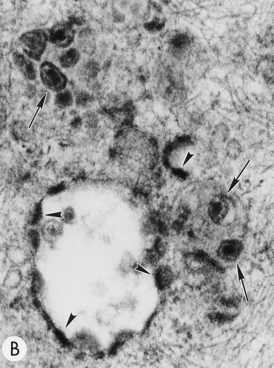
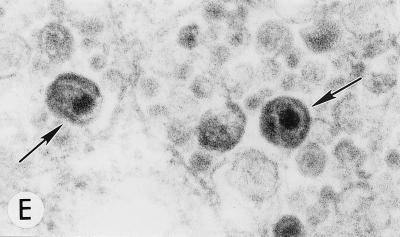
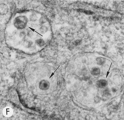
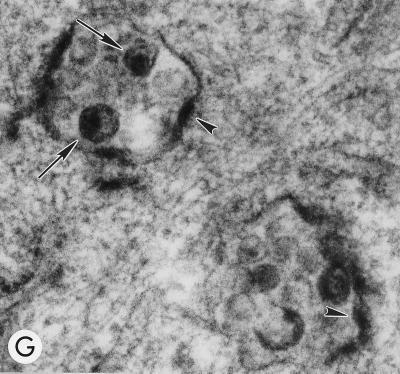
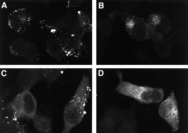
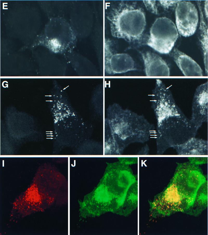
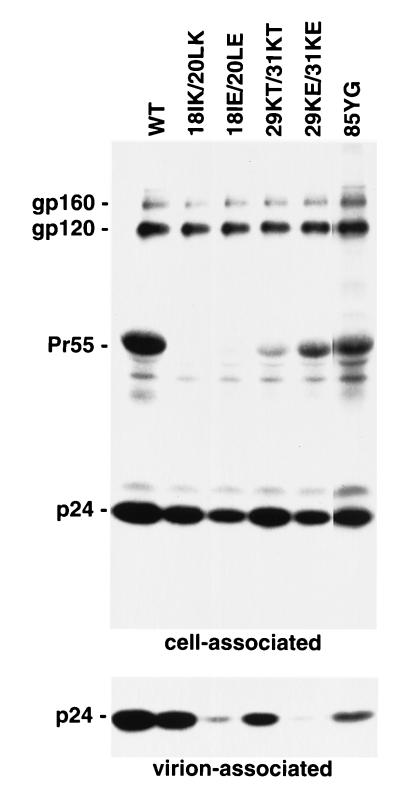
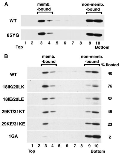
Human immunodeficiency virus type 1 (HIV-1) particle formation and the subsequent initiation of protease-mediated maturation occur predominantly on the plasma membrane. However, the mechanism by which HIV-1 assembly is targeted specifically to the plasma membrane versus intracellular membranes is largely unknown. Previously, we observed that mutations between residues 84 and 88 of the matrix (MA) domain of HIV-1 Gag cause a retargeting of virus particle formation to an intracellular site. In this study, we demonstrate that the mutant virus assembly occurs in the Golgi or in post-Golgi vesicles. These particles undergo core condensation in a protease-dependent manner, indicating that virus maturation can occur not only on the plasma membrane but also in the Golgi or post-Golgi vesicles. The intracellular assembly of mutant particles is dependent on Gag myristylation but is not influenced by p6(Gag) or envelope glycoprotein expression. Previous characterization of viral revertants suggested a functional relationship between the highly basic domain of MA (amino acids 17 to 31) and residues 84 to 88. We now demonstrate that mutations in the highly basic domain also retarget virus particle formation to the Golgi or post-Golgi vesicles. Although the basic domain has been implicated in Gag membrane binding, no correlation was observed between the impact of mutations on membrane binding and Gag targeting, indicating that these two functions of MA are genetically separable. Plasma membrane targeting of Gag proteins with mutations in either the basic domain or between residues 84 and 88 was rescued by coexpression with wild-type Gag; however, the two groups of MA mutants could not rescue each other. We propose that the highly basic domain of MA contains a major determinant of HIV-1 Gag plasma membrane targeting and that mutations between residues 84 and 88 disrupt plasma membrane targeting through an effect on the basic domain.


Figures

FIG. 1
FIG. 1
Evaluation of mutant virus targeting and maturation by EM. HeLa cells transfected with the following molecular clones were fixed and analyzed by EM 2 days posttransfection. Arrows and arrowheads indicate VLPs with condensed cores and budding structures, respectively. (A) WT pNL4-3, showing a cluster of typical mature particles on the cell surface. Magnification, ×83,520. (B) pNL4-3/85YG. Magnification, ×72,210. (C) pNL4-3/87VE/PR. Magnification, ×95,700. (D) pNL4-3KFS/85YG. Magnification, ×89,250. (E) pNL4-3/85YG/p6. Magnification, ×89,250. (F) pNL4-3/29KE/31KE. Magnification, ×94,600. (G) pNL4-3/29KT/31KT. Magnification, ×93,500.
FIG. 1
FIG. 1
Evaluation of mutant virus targeting and maturation by EM. HeLa cells transfected with the following molecular clones were fixed and analyzed by EM 2 days posttransfection. Arrows and arrowheads indicate VLPs with condensed cores and budding structures, respectively. (A) WT pNL4-3, showing a cluster of typical mature particles on the cell surface. Magnification, ×83,520. (B) pNL4-3/85YG. Magnification, ×72,210. (C) pNL4-3/87VE/PR. Magnification, ×95,700. (D) pNL4-3KFS/85YG. Magnification, ×89,250. (E) pNL4-3/85YG/p6. Magnification, ×89,250. (F) pNL4-3/29KE/31KE. Magnification, ×94,600. (G) pNL4-3/29KT/31KT. Magnification, ×93,500.
FIG. 2
FIG. 2
Accumulation of processed mutant Gag in the Golgi and/or post-Golgi vesicles. HeLa cells were transfected with WT pNL4-3 (A and C) or with pNL4-3/85YG (B, D, and E through J) and fixed 2 days posttransfection. After permeabilization, cells were stained with either mouse monoclonal anti-p17 antibody (A and B) or mouse monoclonal anti-p17/55 antibody (C and D) or costained with mouse monoclonal anti-p17 antibody (E, G, and I) and organelle marker rabbit anti-calreticulin antibody (F), RCA60 (H), or WGA (J) and analyzed by confocal microscopy. The same fields are shown for panels E and F, G and H, and I and J. In panels G and H, some vesicles which show double staining for both p17 (MA) and RCA60 are indicated with arrows. Panel I shows 85YG p17 (MA) staining in red and panel J shows WGA staining in green. An overlay of the p17 (MA) and Golgi staining is presented in Panel K; red (MA) and green (Golgi) overlap is visualized as yellow.
FIG. 2
FIG. 2
Accumulation of processed mutant Gag in the Golgi and/or post-Golgi vesicles. HeLa cells were transfected with WT pNL4-3 (A and C) or with pNL4-3/85YG (B, D, and E through J) and fixed 2 days posttransfection. After permeabilization, cells were stained with either mouse monoclonal anti-p17 antibody (A and B) or mouse monoclonal anti-p17/55 antibody (C and D) or costained with mouse monoclonal anti-p17 antibody (E, G, and I) and organelle marker rabbit anti-calreticulin antibody (F), RCA60 (H), or WGA (J) and analyzed by confocal microscopy. The same fields are shown for panels E and F, G and H, and I and J. In panels G and H, some vesicles which show double staining for both p17 (MA) and RCA60 are indicated with arrows. Panel I shows 85YG p17 (MA) staining in red and panel J shows WGA staining in green. An overlay of the p17 (MA) and Golgi staining is presented in Panel K; red (MA) and green (Golgi) overlap is visualized as yellow.
FIG. 3
FIG. 3
WT and mutant virus production. HeLa cells transfected with WT pNL4-3 (WT) or its derivatives containing the indicated MA mutations were metabolically labeled with [35S]Cys. Virions were pelleted by ultracentrifugation. Cell- and virion-associated material was immunoprecipitated with AIDS patient sera and analyzed by sodium dodecyl sulfate-polyacrylamide gel electrophoresis followed by fluorography. The positions of the Env precursor gp160, the mature surface Env glycoprotein gp120, the Gag precursor Pr55Gag, and p24 (CA) are shown.
FIG. 4
FIG. 4
Accumulation of processed Gag in the Golgi and/or post-Golgi vesicles caused by mutations in the MA basic domain. HeLa cells were transfected with WT pNL4-3 (A), pNL4-3/85YG (B), pNL4-3/18IK/20LK (C), pNL4-3/18IE/20LE (D), pNL4-3/29KT/31KT (E), or pNL4-3/29KE/31KE (F). Two days posttransfection, cells were fixed, permeabilized, stained with monoclonal anti-p17 antibody, and analyzed by confocal microscopy.
FIG. 5
FIG. 5
Effects of MA mutations on Gag membrane binding. HeLa cells were transfected with pNL4-3/PR (WT) or its derivatives containing the indicated MA mutations. Postnuclear supernatants were prepared and subjected to equilibrium flotation centrifugation (see Materials and Methods), during which membrane-bound (memb.-bound) materials float to the interface between 10 and 65% sucrose (fractions 3 and 4). Pr55Gag detected by Western blotting is shown. In panel B, the percentage of total Pr55Gag that is membrane bound is indicated on the right.
FIG. 6
FIG. 6
Coexpression of WT and mutant Gag proteins. HeLa cells were transfected at a ratio of 1:1 with various combinations of molecular clones expressing WT, 85YG, or 29KE/31KE Gag tagged with either the HA or the FLAG epitope. Virions were pelleted by ultracentrifugation. Cell and virion lysates were analyzed by Western blotting with anti-HA or anti-FLAG antibodies. Epitope-tagged Pr55Gag is shown.

References

    1. Adachi A, Gendelman H E, Koenig S, Folks T, Willey R, Rabson A, Martin M A. Production of acquired immunodeficiency syndrome-associated retrovirus in human and nonhuman cells transfected with an infectious molecular clone. J Virol. 1986;59:284–291. - PMC - PubMed
    1. Aloia R C, Tian H, Jensen F C. Lipid composition and fluidity of the human immunodeficiency virus envelope and host cell plasma membranes. Proc Natl Acad Sci USA. 1993;90:5181–5185. - PMC - PubMed
    1. Brown D A, London E. Structure of detergent-resistant membrane domains: does phase separation occur in biological membranes? Biochem Biophys Res Commun. 1997;240:1–7. - PubMed
    1. Bryant M, Ratner L. Myristoylation-dependent replication and assembly of human immunodeficiency virus 1. Proc Natl Acad Sci USA. 1990;87:523–527. - PMC - PubMed
    1. Cannon P M, Matthews S, Clark N, Byles E D, Iourin O, Hockley D J, Kingsman S M, Kingsman A J. Structure-function studies of the human immunodeficiency virus type 1 matrix protein, p17. J Virol. 1997;71:3474–3483. - PMC - PubMed

MeSH terms

Substances