Case Reports
. 2000 Mar;84(3):251-8.
doi: 10.1136/bjo.84.3.251.

Radiological and clinicopathological features of orbital xanthogranuloma

Affiliations
Case Reports

Radiological and clinicopathological features of orbital xanthogranuloma

K A Miszkiel et al. Br J Ophthalmol. 2000 Mar.

Abstract

Background: Orbital xanthogranuloma, a diagnosis confirmed histologically, occurs rarely in adults and children. With its characteristic macroscopic appearance the adult form may be associated with a spectrum of biochemical and haematological abnormalities including lymphoproliferative malignancies.

Method: The clinicopathological features and imaging appearances on computed tomography and magnetic resonance imaging of this condition are described in eight adults and a child.

Results: Radiological evidence of proptosis was present in seven patients. In all nine patients an abnormal infiltrative soft tissue mass was seen, with increased fat in six cases. All patients had associated enlargement of extraocular muscles suggestive of infiltration and five had lacrimal gland involvement. Encasement of the optic nerve, bone destruction, and intracranial extension was present only in the child with juvenile xanthogranuloma. Haematological and/or biochemical abnormalities were detected in seven patients and seven patients had other systemic diseases which were considered to have an immune basis. One patient subsequently developed non-Hodgkin's lymphoma.

Conclusion: The investigation and management of orbital xanthogranulomas requires a multidisciplinary approach even though the diagnosis may be suspected clinically. Imaging delineates the extent of disease and involvement of local structures and may influence the differential diagnosis. The juvenile form may be more locally aggressive, causing bone destruction with consequent intracranial extension.


Figures
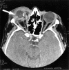
Figure 1
Figure 1
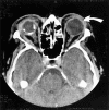
Contrast enhanced axial computed tomogram demonstrating a poorly defined, infiltrative soft tissue and fat containing mass in the right preseptal soft tissues (arrow) with extension around the small deformed calcified globe. There is proptosis and enlargement of the right medial rectus muscle.
Figure 2
Figure 2
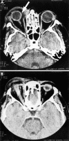
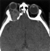
Follow up computed tomogram 11 months later after treatment with radiotherapy and chlorambucil, showed some resolution of changes affecting the right orbit, but progressive infiltration of the left preseptal soft tissues by a similar process (curved arrow).
Figure 3
Figure 3
Biopsy sections through the skin show extensive infiltration of the dermis by xanthoma cells and also occasional areas of lymphoid infiltration, (A) haematoxylin and eosin and (B) oil red O, frozen for fat; original magnification ×100.
Figure 4
Figure 4
Unenhanced axial computed tomogram showing a mixed soft tissue density mass affecting the upper lid (arrow) in (A), and extending posteriorly above the globe, with enlargement and some irregularity of the margins of the superior rectus muscle (B). Again there is marked proptosis.
Figure 5
Figure 5
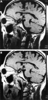
Parasagittal T-1 weighted unenhanced image through the right orbit demonstrates the fat and soft tissue density mass affecting the upper lid and extending posteriorly above the superior rectus muscle (curved arrow) in (A). After contrast there is enhancement of the soft tissue components of the mass (B).
Figure 6
Figure 6
Biopsy demonstrating perivascular infiltration of the orbital tissue by foamy macrophages with occasional lymphocytes. (A) Haematoxylin and eosin with a similar high power field stained by oil red O (B). The original magnification of both images is ×400.
Figure 7
Figure 7
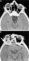
Unenhanced computed tomogram showing marked right proptosis due to a large ovoid soft tissue mass posterolaterally within the orbit (A), which abuts the globe superiorly (B). Some sclerosis of the lateral orbital margin is seen (arrow) in (B).
Figure 8
Figure 8
Six months later the mass has increased in size, appears more heterogeneous in texture, and there is now definite bony destruction and intracranial extension.

References

    1. Am J Ophthalmol. 1966 May;61(5 Pt 2):1299-306 - PubMed
    1. Trans Am Ophthalmol Soc. 1960;58:59-74 - PubMed
    1. Australas J Dermatol. 1980 Aug;21(2):85-8 - PubMed
    1. Am J Ophthalmol. 1984 Feb;97(2):173-83 - PubMed
    1. Australas Radiol. 1984 Nov;28(4):305-10 - PubMed

Publication types