. 2000 Mar;84(3):311-7.
doi: 10.1136/bjo.84.3.311.

Age related changes in the non-collagenous components of the extracellular matrix of the human lamina cribrosa

Affiliations

Age related changes in the non-collagenous components of the extracellular matrix of the human lamina cribrosa

J Albon et al. Br J Ophthalmol. 2000 Mar.

Abstract

Aims: To investigate age related alterations in the non-collagenous components of the human lamina cribrosa.

Methods: Fibronectin, elastin, and glial fibrillary acidic protein (GFAP) staining were assessed in young and old laminae cribrosae. An age range (7 days to 96 years) of human laminae cribrosae were analysed for lipid content (n=9), cellularity (n=28), total sulphated glycosaminoglycans (n=28), elastin content (n=9), and water content (n=56), using chloroform-methanol extraction, fluorimetry, the dimethylmethylene blue assay, and ion exchange chromatography, respectively.

Results: Qualitatively, an increase in elastin and a decrease in fibronectin and GFAP were demonstrated when young tissue was compared with the elderly. Biochemical analysis of the ageing human lamina cribrosa demonstrated that elastin content increased from 8% to 28% dry tissue weight, total sulphated glycosaminoglycans decreased, and lipid content decreased from 45% to 25%. There were no significant changes in total cellularity or water content.

Conclusion: These alterations in composition may be indicative of the metabolic state of the lamina cribrosa as it ages, and may contribute to changes in mechanical integrity. Such changes may be implicated in the susceptibility of the elderly lamina cribrosa and also its response to glaucomatous optic neuropathy.


Figures
None
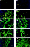
Figures 1-3(1) Cross sections of human lamina cribrosa: demonstrating the distribution of elastin staining in a 16 year old (1a and 1b) (magnification ×140) and an 86 year old (1c and 1d) (magnification ×230). Positive localisation of elastin was demonstrated in (1a) the walls of the sclera and in the extrafascicular matrix of the young lamina cribrosa, and (1b) the elastic lamina of the central retinal artery; very faint staining was also observed in the walls of the central retinal vessels. (1c) Much denser staining within the sclera and extrafascicular matrix and (1d) the sheaths of the central retinal vessels was demonstrated in the 86 year old. (2) Cross sections of lamina cribrosa, immunolocalised for the glycoprotein fibronectin in tissue aged 16 years (2a and 2b) and 86 years (2c and 2d). Positive staining was demonstrated in the extrafascicular matrix (2a and 2c) (magnification ×140) and throughout the walls of the central retinal vessels, although staining was much weaker in the 86 year old (magnification ×230). (3) Immunolocalisation of glial fibrillary acidic protein (GFAP) in cross sections of lamina cribrosa (3a) 16 year old lamina cribrosa (magnification ×140): GFAP labelling was demonstrated at the region of insertion of the lamina cribrosa into the sclera (Sc), and throughout the nerve fibre bundles (nb). (3b) 78 year old lamina cribrosa (magnification ×140): GFAP labelling followed the same distribution as that described in the young lamina, but staining was much weaker. Note the high amount of autofluorescence.
Figure 4
Figure 4
Water content of the ageing human lamina cribrosa. Percentage water content of the human lamina cribrosa, calculated as the difference between wet weight and dry weight after freeze drying, remained constant throughout life. Spearman's rank correlation rs=0.23, not significant at p=0.039, n=56).
Figure 5
Figure 5
Lipid content in the ageing human lamina cribrosa. Percentage lipid was calculated in nine groups of laminae cribrosae aged between 1 and 89 years of age. The dry weight of each age group was measured after extraction of lipid from each group in 3:1 chloroform:methanol. The difference between this value and that of the total dry weight was expressed as a percentage of total dry tissue weight to provide a value for lipid content in each age category. Lipid content decreased linearly from 42% to 24%. (Line fitted is a first order polynomial regression line r=0.86.)
Figure 6
Figure 6
DNA content of human lamina cribrosa. DNA content was measured in an aliquot taken from papain digested laminae cribrosae, using Hoechst 33256. Fluorescence of DNA-Hoechst complex was detected at excitation/emission wavelengths of 328 nm/457 nm using a Perkin-Elmer fluorimeter. DNA content decreased with age, not significant at p=0.291. Spearman's rank correlation rs= −0.21, n=28.
Figure 7
Figure 7
Sulphated glycosaminoglycans in the human lamina cribrosa. Sulphated GAGs analysis of papain digests of paired laminae cribrosae was carried out using the dimethylmethylene blue dye binding assay at an absorbance of 540 nm. Sulphated GAGs content of the human lamina cribrosa decreased with age. (Spearman's rank correlation rs= −0.45, p=0.017, fitted line is a regression line, r=0.75, n=28).
Figure 8
Figure 8
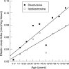
Elastin cross links in the human lamina cribrosa. The elastin cross links, desmosine and isodesmosine, were analysed using ion exchange chromatography, with ninhydrin detection.21 Both cross links significantly increased with age. (Lines fitted are first order polynomial regression lines with correlation coefficients r=0.87 and r=0.85, for desmosine and isodesmosine respectively.)
Figure 9
Figure 9
Elastin content of the human lamina cribrosa. Elastin content was estimated from the values of desmosine, and expressed as a percentage of total dry tissue weight. Percentage elastin increased significantly with age from 7% to 28%. (Fitted line is a second order polynomial regression fit, r=0.87.)
Figure 10
Figure 10
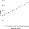
Collagen content of the human lamina cribrosa. Collagen content expressed as a percentage of total dry tissue weight increased from 24% to 47%.

References

    1. Curr Eye Res. 1993 Aug;12(8):685-92 - PubMed
    1. Ann Med. 1993 Aug;25(4):309-15 - PubMed
    1. Surv Ophthalmol. 1994 Jul-Aug;39(1):23-42 - PubMed
    1. Br J Ophthalmol. 1995 Apr;79(4):368-75 - PubMed
    1. Glia. 1997 Jul;20(3):262-73 - PubMed

Publication types