. 2000 Mar;84(3):318-23.
doi: 10.1136/bjo.84.3.318.

Age related compliance of the lamina cribrosa in human eyes

Affiliations

Age related compliance of the lamina cribrosa in human eyes

J Albon et al. Br J Ophthalmol. 2000 Mar.

Abstract

Aims: To investigate changes in the mechanical compliance of ex vivo human lamina cribrosa with age.

Methods: A laser scanning confocal microscope was used to image the surface of the fluorescently labelled lamina cribrosa in cadaver eyes. A method was developed to determine changes in the volume and strain of the lamina cribrosa created by increases in pressure. The ability of the lamina cribrosa to reverse its deformation on removal of pressure was also measured.

Results: Volume and strain measurements both demonstrated that the lamina cribrosa increased in stiffness with age and the level of pressure applied. The ability of the lamina cribrosa to regain its original shape and size on removal of pressure appeared to decrease with age, demonstrating an age related decrease in resilience of the lamina cribrosa.

Conclusions: The mechanical compliance of the human lamina cribrosa decreased with age. Misalignment of compliant cribriform plates in a young eye may exert a lesser stress on nerve axons, than that exerted by the rigid plates of an elderly lamina cribrosa. The resilience of the lamina cribrosa also decreased with age, suggesting an increased susceptibility to plastic flow and permanent deformation. Such changes may be of importance in the explanation of age related optic neuropathy in primary open angle glaucoma.


Figures

Figure 1
Figure 1
Schematic diagram of apparatus used to measure the change in dimensions of the lamina cribrosa on application and removal of pressure.
Figure 2
Figure 2
A typical xy scan demonstrating the fluorescently labelled anterior surface of the human lamina cribrosa, imaged using a Bio-Rad MRC-500 confocal laser scanning microscope. An average of 35 scans using Kalman were imaged for each xy scan.
Figure 3
Figure 3
A confocal xz scan demonstrating the deformation of a lamina cribrosa at atmospheric pressure (solid line) and after the pressure has been increased by 43 mm Hg (broken line). Settings included: start scan 300 µm above sample surface, incremental step of 25 (2.5), motor step drive of 400.
Figure 4
Figure 4
Pressure-volume curves of a young (9 year old: circles) and old (86 year old: squares) lamina cribrosa demonstrating their non-linearity and the difference in gradients. Solid line denotes loading curve as pressure was increased and the broken line denotes the unloading curve as pressure was decreased. Residual volume is defined as the difference between the initial volume and the volume after the removal of applied pressure.
Figure 5
Figure 5
Reversibility of the lamina cribrosa versus age. Reversibility of the lamina cribrosa on reduction of pressure was expressed as the volume recovered as a percentage of total volume change. Line shown is a fitted second order polynomial regression y=81.089 − 0.89614x + 4.153e−3x2 r2=0.776.
Figure 6
Figure 6
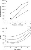
Incremental gradients (dP/dS, that is to say the change in pressure relative to the change in strain) were calculated for each pressure point from a polynomial regression curve through the loading curve of each lamina cribrosa. The incremental gradient defined the changes in the mechanical compliance of the lamina cribrosa with age, as pressure was increased. (A) Incremental gradients of the pressure-strain curves for three of the 10 eyes analysed: 7 (squares), 44 (triangles) and 86 (circles) year old, versus the pressure applied to produce each strain. The incremental gradient increased with age and pressure, indicating a stiffening of the lamina cribrosa. (B) Incremental gradients dP/dS versus age for given pressure increments, indicating a rapid increase in stiffness of the lamina cribrosa after 40-50 years of age. Lines show second order polynomial regression. The correlation values (r) vary between 0.65 and 0.75 for all curves shown.
Figure 7
Figure 7
Strains in two directions across the lamina cribrosa S1 and S2: the strain in one of the two directions was always greater in one dimension.

References

    1. Invest Ophthalmol Vis Sci. 1979 Feb;18(2):166-71 - PubMed
    1. Curr Eye Res. 1998 Sep;17(9):903-16 - PubMed
    1. Arch Ophthalmol. 1981 Jan;99(1):137-43 - PubMed
    1. J Opt Soc Am. 1981 Apr;71(4):499-501 - PubMed
    1. Arch Ophthalmol. 1981 Dec;99(12):2159-62 - PubMed

Publication types