. 2000 Feb 21;191(4):593-602.
doi: 10.1084/jem.191.4.593.

Helicobacter pylori CagA protein can be tyrosine phosphorylated in gastric epithelial cells

Affiliations

Helicobacter pylori CagA protein can be tyrosine phosphorylated in gastric epithelial cells

M Asahi et al. J Exp Med. .

Abstract

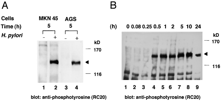
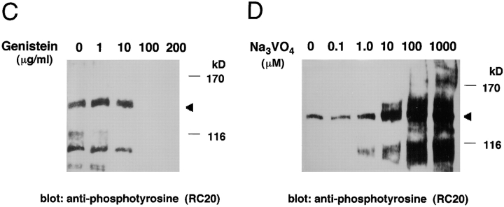
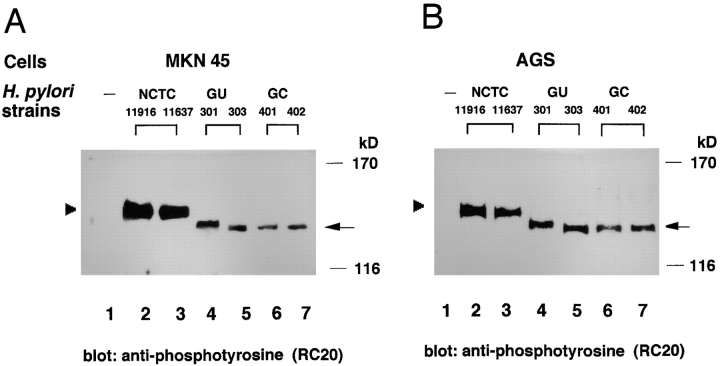
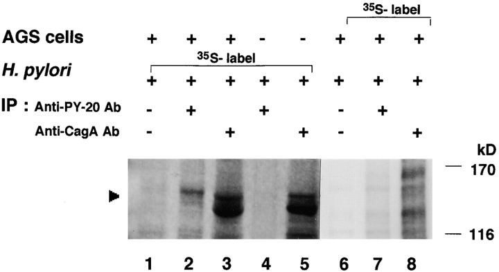
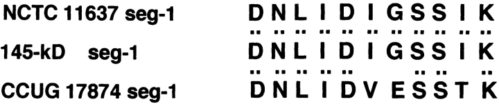
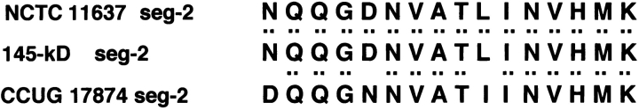
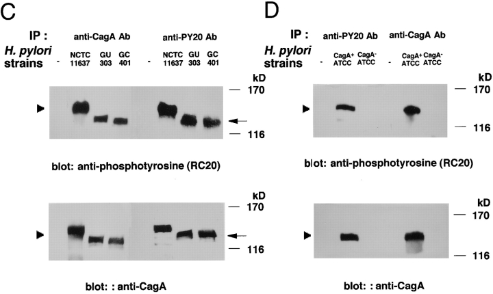
Attachment of Helicobacter pylori to gastric epithelial cells induces various cellular responses, including the tyrosine phosphorylation of an unknown 145-kD protein and interleukin 8 production. Here we show that this 145-kD protein is the cagA product of H. pylori, an immunodominant, cytotoxin-associated antigen. Epithelial cells infected with various H. pylori clinical isolates resulted in generation of tyrosine-phosphorylated proteins ranging from 130 to 145 kD in size that were also induced in vitro by mixing host cell lysate with bacterial lysate. When epithelial cells were infected with [(35)S]methionine-labeled H. pylori, a radioactive 145-kD protein was detected in the immunoprecipitates with antiphosphotyrosine antibody or anti-CagA (cytotoxin-associated gene A) antibody. Consistently, the 145-kD protein recognized by the anti-CagA and antiphosphotyrosine antibodies was induced in epithelial cells after infection of wild-type H. pylori but not the cagA::Km mutant. Furthermore, the amino acid sequence of the phosphorylated 145-kD protein induced by H. pylori infection was identical to the H. pylori CagA sequence. These results reveal that the tyrosine-phosphorylated 145-kD protein is H. pylori CagA protein, which may be delivered from attached bacteria into the host cytoplasm. The identification of the tyrosine-phosphorylated protein will thus provide further insights into understanding the precise roles of CagA protein in H. pylori pathogenesis.


Figures

Figure 1
Figure 1
Immunoblot analysis of gastric epithelial cells infected with H. pylori by antiphosphotyrosine antibody RC-20. (A) Tyrosine phosphorylation of a 145-kD protein in MKN45 or AGS cells induced by H. pylori NCTC11637 infection. Lanes 1 and 2, lysates of uninfected MKN45 or MKN45 infected with H. pylori for 5 h; lanes 3 and 4, lysates of uninfected AGS or AGS infected with H. pylori for 5 h. Arrowhead indicates the 145-kD protein. (B) Time course for the expression of protein tyrosine phosphorylation in MKN45 cells infected with H. pylori NCTC11637. Uninfected (lane 1) or infected (lanes 2–9) MKN45 cells were examined at the indicated times (h) after bacterial infection. Effect of genistein (C) or vanadate (D) on the 145-kD protein tyrosine phosphorylation in MKN45 cells infected with H. pylori NCTC11637. Cells were treated with various concentrations of genistein or vanadate for 30 min at 37°C before the infection. The epithelial cells infected with H. pylori for 5 h were sampled at the indicated concentration, and the protein tyrosine phosphorylation in the cell lysates was examined by the same methods as in A.
Figure 1
Figure 1
Immunoblot analysis of gastric epithelial cells infected with H. pylori by antiphosphotyrosine antibody RC-20. (A) Tyrosine phosphorylation of a 145-kD protein in MKN45 or AGS cells induced by H. pylori NCTC11637 infection. Lanes 1 and 2, lysates of uninfected MKN45 or MKN45 infected with H. pylori for 5 h; lanes 3 and 4, lysates of uninfected AGS or AGS infected with H. pylori for 5 h. Arrowhead indicates the 145-kD protein. (B) Time course for the expression of protein tyrosine phosphorylation in MKN45 cells infected with H. pylori NCTC11637. Uninfected (lane 1) or infected (lanes 2–9) MKN45 cells were examined at the indicated times (h) after bacterial infection. Effect of genistein (C) or vanadate (D) on the 145-kD protein tyrosine phosphorylation in MKN45 cells infected with H. pylori NCTC11637. Cells were treated with various concentrations of genistein or vanadate for 30 min at 37°C before the infection. The epithelial cells infected with H. pylori for 5 h were sampled at the indicated concentration, and the protein tyrosine phosphorylation in the cell lysates was examined by the same methods as in A.
Figure 2
Figure 2
Diversity of tyrosine-phosphorylated large proteins induced in epithelial cells infected with various H. pylori strains. Protein tyrosine phosphorylation in MKN45 (A) and AGS cells (B) infected with various clinical isolates of H. pylori for 5 h. Lane 1, uninfected control; lanes 2–7 represent protein tyrosine phosphorylation induced in epithelial cells infected with NCTC11916, NCTC11637, GU301, GU303, GC401, and GC402, respectively. Arrowhead indicates the tyrosine-phosphorylated 145-kD protein; arrow indicates the phosphorylated large proteins in epithelial cells infected with various clinical isolates.
Figure 3
Figure 3
In vitro tyrosine phosphorylation of the 145-kD protein. (A) Tyrosine phosphorylation of a 145-kD protein induced by a combination of H. pylori NCTC11637 and MKN45 (or AGS) cell lysates. Epithelial cell lysates with or without bacterial lysates were added to a phosphorylation reaction buffer and incubated for 10 min at 30°C. The 145-kD protein was detected by immunoblotting with antiphosphotyrosine antibody RC-20. (B) Tyrosine phosphorylation of a large protein induced by the combination of various H. pylori strains and MKN45 cell lysates. Lanes 1–9: control (without H. pylori lysate), NCTC11637, GU301, GU303, GU304, GU305, GU306, GC401, and GC402, respectively. Arrowhead and arrow indicate the tyrosine-phosphorylated 145-kD and other proteins, respectively.
Figure 4
Figure 4
Demonstration of the tyrosine-phosphorylated 145-kD protein derived from H. pylori. AGS cells were infected with [35S]methionine-labeled or nonlabeled H. pylori for 5 h at 37°C, and the cell lysates prepared with 1% Triton X-100 were immunoprecipitated by an antiphosphotyrosine mAb PY-20 or an anti-CagA polyclonal antibody. The precipitated proteins were separated by SDS-PAGE and analyzed using a radioanalytic imaging system. Arrowhead indicates the 145-kD protein. Lanes 1–3, AGS infected with [35S]methionine-labeled H. pylori; lanes 4 and 5, [35S]methionine-labeled H. pylori alone; lanes 6–8, [35S]methionine-labeled AGS infected with nonlabeled H. pylori; lanes 1 and 6, the 1% Triton X-100–soluble fraction precipitated by control normal IgG; lanes 2, 4, and 7, the 1% Triton X-100–soluble fraction precipitated by antiphosphotyrosine mAb PY-20; and lanes 3, 5, and 8, the 1% Triton X-100–soluble fraction precipitated by anti-CagA polyclonal antibody. Note that the [35S]methionine-labeled band being lower than that of 145-kD protein in lanes 3 and 5 may be bacterial protein(s) cross-reacted with the polyclonal anti-CagA antibody.
Figure 5
Figure 5
Amino acid sequences of the tyrosine-phosphorylated 145-kD protein. Amino acid sequences of the three internal segments of the tyrosine-phosphorylated 145-kD protein (145-kD seg-1, -2, and -3) from in vitro sample (the reactant of NCTC11637 and MKN45 cell lysates incubated in the phosphorylation reaction buffer in vitro), the amino acid sequences deduced from nucleotide sequences of the DNA segments encoding sequences 1, 2, and 3 from NCTC11637 (NCTC 11637 seg-1, -2, and -3), or the amino acid sequences deduced from the nucleotide sequences of the corresponding cagA sequence from CCUG17874 (CCUG 17874 seg-1, -2, and -3; reference 11).
Figure 5
Figure 5
Amino acid sequences of the tyrosine-phosphorylated 145-kD protein. Amino acid sequences of the three internal segments of the tyrosine-phosphorylated 145-kD protein (145-kD seg-1, -2, and -3) from in vitro sample (the reactant of NCTC11637 and MKN45 cell lysates incubated in the phosphorylation reaction buffer in vitro), the amino acid sequences deduced from nucleotide sequences of the DNA segments encoding sequences 1, 2, and 3 from NCTC11637 (NCTC 11637 seg-1, -2, and -3), or the amino acid sequences deduced from the nucleotide sequences of the corresponding cagA sequence from CCUG17874 (CCUG 17874 seg-1, -2, and -3; reference 11).
Figure 6
Figure 6
Tyrosine phosphorylation of CagA protein induced in H. pylori–infected epithelial cells. (A) Detection of the tyrosine phosphorylation of CagA protein in AGS cells induced by H. pylori infection. AGS cells infected with H. pylori NCTC11637 at indicated times after infection were lysed in a 1% Triton X-100 lysis buffer, and the soluble fraction was immunoprecipitated with an anti-CagA polyclonal antibody. The precipitates were separated by SDS-PAGE and then immunoblotted with antiphosphotyrosine antibody RC-20 or anti-CagA polyclonal antibody. (B) Absence of tyrosine-phosphorylated CagA protein in the culture supernatants. The culture supernatants used for the AGS cells infected with H. pylori NCTC11637 at the indicated times were subjected to the same immunoblot analysis as in A. The rightmost lane represents the tyrosine phosphorylation of CagA protein in AGS cells induced by H. pylori 10 h after infection. (C) Tyrosine phosphorylation of CagA protein in AGS cells induced by infection with H. pylori strains NCTC11637, GU303, and GC401. The leftmost lane shows noninfected control, and the second lane on the left shows NCTC11637 control. Top panel: tyrosine-phosphorylated CagA proteins detected in AGS cells infected with H. pylori; bottom panel: the same blot reprobed with the anti-CagA polyclonal antibody. (D) Tyrosine phosphorylation of CagA protein in AGS cells induced by infection with ATCC43579 and the cagA::Km mutant. Arrowhead indicates the 145-kD protein.
Figure 6
Figure 6
Tyrosine phosphorylation of CagA protein induced in H. pylori–infected epithelial cells. (A) Detection of the tyrosine phosphorylation of CagA protein in AGS cells induced by H. pylori infection. AGS cells infected with H. pylori NCTC11637 at indicated times after infection were lysed in a 1% Triton X-100 lysis buffer, and the soluble fraction was immunoprecipitated with an anti-CagA polyclonal antibody. The precipitates were separated by SDS-PAGE and then immunoblotted with antiphosphotyrosine antibody RC-20 or anti-CagA polyclonal antibody. (B) Absence of tyrosine-phosphorylated CagA protein in the culture supernatants. The culture supernatants used for the AGS cells infected with H. pylori NCTC11637 at the indicated times were subjected to the same immunoblot analysis as in A. The rightmost lane represents the tyrosine phosphorylation of CagA protein in AGS cells induced by H. pylori 10 h after infection. (C) Tyrosine phosphorylation of CagA protein in AGS cells induced by infection with H. pylori strains NCTC11637, GU303, and GC401. The leftmost lane shows noninfected control, and the second lane on the left shows NCTC11637 control. Top panel: tyrosine-phosphorylated CagA proteins detected in AGS cells infected with H. pylori; bottom panel: the same blot reprobed with the anti-CagA polyclonal antibody. (D) Tyrosine phosphorylation of CagA protein in AGS cells induced by infection with ATCC43579 and the cagA::Km mutant. Arrowhead indicates the 145-kD protein.

Comment in

References

    1. Marshall B.J., Warren J.R. Unidentified curved bacilli in the stomach of patients with gastritis and peptic ulceration. Lancet. 1984;1:1311–1315. - PubMed
    1. Goodwin C.S., Armstrong J.A., Marshall B.J. Campylobacter pyloridis, gastritis, and peptic ulceration. J. Clin. Pathol. 1986;39:353–365. - PMC - PubMed
    1. Blaser M.J. Helicobacter pylori and the pathogenesis of gastroduodenal inflammation. J. Infect. Dis. 1990;161:626–633. - PubMed
    1. Parsonnet J., Friedman G.D., Vandersteen D.P., Chang Y., Vogelman J.H., Orentreich N., Sibley R.K. Helicobacter pylori infection and the risk of gastric carcinoma. N. Engl. J. Med. 1991;325:1127–1131. - PubMed
    1. Parsonnet J., Hansen S., Rodriguez L., Gelb A.B., Warnke R.A., Jellum E., Orentreich N., Vogelman J.H., Friedman G.D. Helicobacter pylori infection and gastric lymphoma. N. Engl. J. Med. 1994;330:1267–1271. - PubMed

Publication types

MeSH terms