. 2000 Feb 21;191(4):631-40.
doi: 10.1084/jem.191.4.631.

Promyelocytic leukemia protein (PML) and Daxx participate in a novel nuclear pathway for apoptosis

Affiliations

Promyelocytic leukemia protein (PML) and Daxx participate in a novel nuclear pathway for apoptosis

S Zhong et al. J Exp Med. .

Abstract

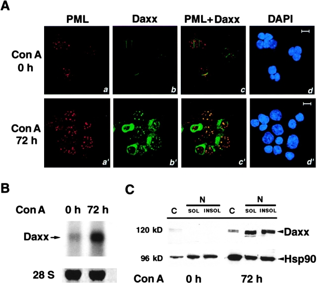
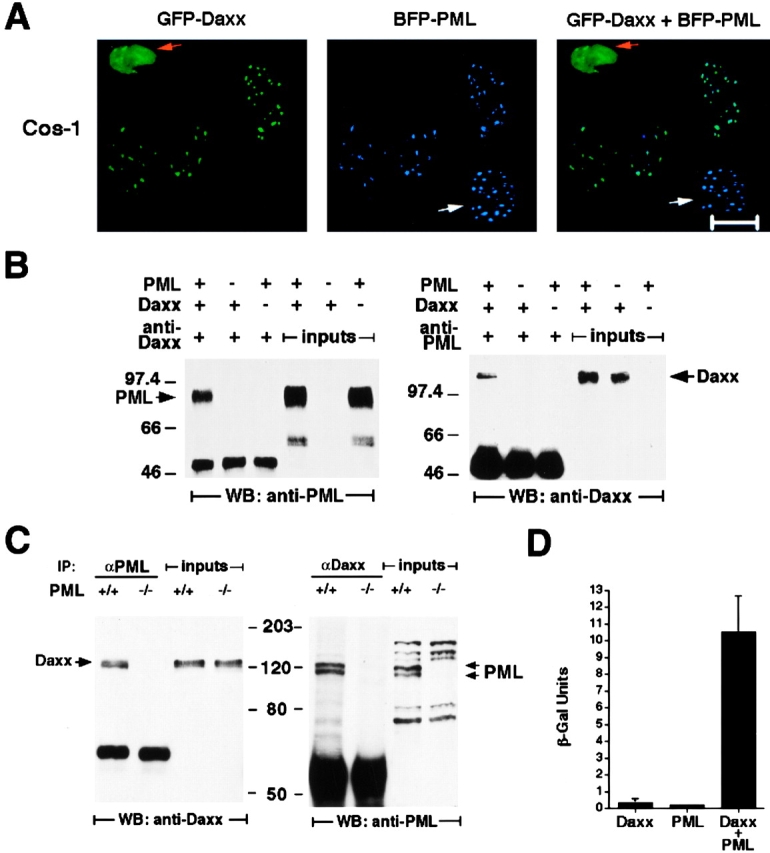
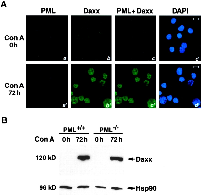
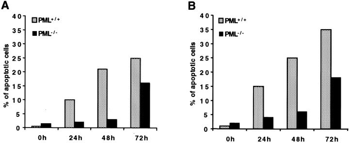
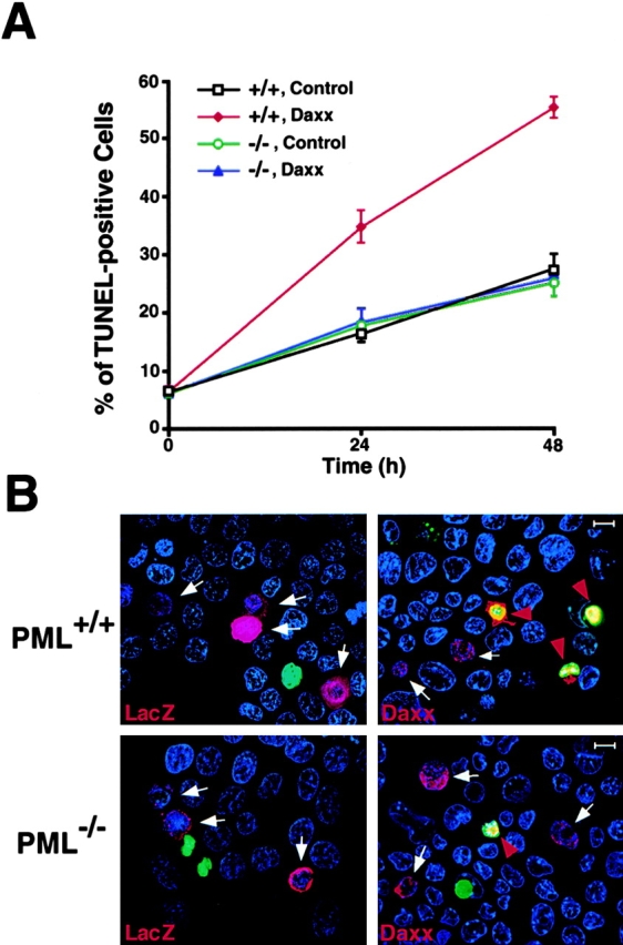
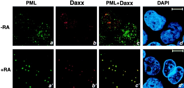
The promyelocytic leukemia protein (PML) gene of acute promyelocytic leukemia (APL) encodes a cell growth and tumor suppressor essential for multiple apoptotic signals. Daxx was identified as a molecule important for the cytoplasmic transduction of the Fas proapoptotic stimulus. Here, we show that upon mitogenic activation of mature splenic lymphocytes, Daxx is dramatically upregulated and accumulates in the PML nuclear body (NB) where PML and Daxx physically interact. In the absence of PML, Daxx acquires a dispersed nuclear pattern, and activation-induced cell death of splenocytes is profoundly impaired. PML inactivation results in the complete abrogation of the Daxx proapoptotic ability. In APL cells, Daxx is delocalized from the NB. Upon retinoic acid treatment, which induces disease remission in APL, Daxx relocalizes to the PML NBs. These results indicate that PML and Daxx cooperate in a novel NB-dependent pathway for apoptosis and shed new light in the role of PML in tumor suppression.


Figures

Figure 1
Figure 1
Con A activation of splenocytes induces Daxx expression. (A) Daxx localization in NBs is induced upon Con A activation. PML+/+ splenocytes, before and after Con A activation, were immunostained with anti-PML and anti-Daxx antibodies. Both Daxx and PML display a strong staining upon Con A activation. Single staining (a, b, a′, and b′) and superimposed images (c and c′) are shown. The nuclei were visualized by DAPI staining (d and d′). Representative confocal pictures are shown. Bar, 10 μM. (B) Upregulation of Daxx mRNA expression upon Con A treatment. Northern blot analysis was performed on total RNA from PML+/+ primary splenocytes, before and after 72 h of Con A activation using a Daxx cDNA probe, which detected a single 2.6-kb band. Normalization with 28S RNA is also shown. (C) Analysis of Daxx protein levels in cytosolic (C) and nuclear NP-40 (N)–soluble (SOL) and insoluble fractions (INSOL). Levels of heat shock protein 90 (Hsp90) were measured as a control of equal loading.
Figure 2
Figure 2
Daxx and PML colocalize in NBs and physically interact. (A) Colocalization of transfected Daxx and PML. Cos-1 cells cotransfected with GFP-Daxx and BFP-PML were analyzed by confocal microscopy. Green dots (GFP) are Daxx specific, blue dots (BFP) are PML specific, and turquoise dots (GFP plus BFP) represent colocalizing proteins. Orange and white arrows indicate cells transfected with Daxx only and with PML only, respectively. Representative pictures are shown. Bar, 10 μM. (B) Coimmunoprecipitation of transfected Daxx and PML in Cos-1 cells. The left panel shows that PML is specifically detected in the Daxx immunoprecipitant, and the right panel shows that Daxx is coimmunoprecipitated with PML. The whole cell lysates (inputs) used for the immunoprecipitation are also included to display the migration of specific immunoreactive species. The anti–human PML antibody used for the coimmunoprecipitation analysis does not cross-react with the endogenous monkey PML. However, Cos-1 cells do express PML as revealed by Northern blot analysis. (C) Coimmunoprecipitation of endogenous Daxx and PML in Con A–activated PML+/+ and PML−/− splenocytes. The left panel shows that Daxx is specifically detected in the PML immunoprecipitant, and the right panel shows that PML is coimmunoprecipitated with Daxx. The migration of specific immunoreactive species detected in the whole cell lysates is also shown. As expected, the endogenous Daxx was coimmunoprecipitated along with PML in PML+/+ but not in PML−/− cells. (D) Yeast two-hybrid assay between the full-length Daxx and PML. Yeast strain Y187 was transformed with pAS2-1-Daxx and/or pACT-2-PML. Three to four colonies per transformation were grown in liquid culture, and the β-galactosidase expression (β-Gal) was determined using ONPG as substrate. The expression level of β-galactosidase measured by quantitative liquid assays in the cotransformed colonies was ∼50-fold higher than that in the single-transformed colonies.
Figure 3
Figure 3
PML is required for the NB localization of Daxx. (A) Immunofluorescence analysis of Daxx in PML−/− splenocytes. Splenocytes, before and after Con A activation, were immunostained with anti-PML and anti-Daxx antibodies. Single staining (a, b, a′, and b′) and superimposed images (c and c′) are shown. The nuclei were visualized by DAPI staining (d and d′). Representative confocal pictures are shown. Bar, 10 μM. (B) Western blot analysis of nuclear extract from PML+/+ and PML−/− splenocytes, before and after Con A activation. Hsp90 protein levels are shown as a control of equal loading.
Figure 4
Figure 4
Daxx is selectively induced in B lymphocytes. PML+/+ and PML−/− splenocytes were treated with Con A and at 72 h B and T subpopulations were obtained (Materials and Methods). Daxx expression levels at 0 and 72 h were measured in B and T cells by Western blot with the polyclonal anti-Daxx antibody. The differential expression of Daxx is also visible in extracts from untreated B and T cells upon longer exposure (not shown). Hsp90 was used as a normalization marker for protein input.
Figure 5
Figure 5
Daxx induction correlates with B cell apoptosis. PML+/+ and PML−/− splenocytes were treated with Con A. At 0, 24, 48, and 72 h after stimulation, B lymphocytes were isolated (see Materials and Methods) and scored for apoptosis by in situ TUNEL assay (A) or by cytometric analysis of subdiploid DNA content (B).
Figure 6
Figure 6
PML is required for the ability of Daxx to induce apoptosis. (A) Time course of apoptosis in PML+/+ and PML−/− keratinocytes transfected with His(6×)-Daxx or, as control, His(6×)-LacZ. Apoptotic cells were scored by TUNEL as described in B. The values are expressed as mean percentage of apoptosis ± SD (n = 3). (B) TUNEL staining of PML+/+ (top) and PML−/− (bottom) keratinocytes 24 h after transfection. Transfected cells were recognized by immunofluorescence staining with an anti-His mAb. His(6×)-positive, red; TUNEL-positive, green; DAPI, blue. White arrows point to His(6×)-positive nonapoptotic (TUNEL-negative) cells; red arrowheads point to His(6×)-positive apoptotic cells (yellow). The pictures were taken with an Olympus fluorescence microscope.
Figure 7
Figure 7
Daxx is delocalized in the NB4 cells and relocalized to the NB upon RA treatment. NB4 cells were grown with or without 1 μM RA for 24 h before harvest. Cells were immunostained with a polyclonal anti-Daxx antibody and an anti-PML mAb. Single staining (a, b, a′, and b′), superimposed images (c and c′), and DAPI staining (d and d′) are shown. Representative confocal pictures are shown. Bar, 10 μM.

References

    1. Pandolfi P.P., Grignani F., Alcalay M., Mencarelli A., Biondi A., Lo Coco F., Pelicci P.G. Structure and origin of the acute promyelocytic leukemia myl/RARα cDNA and characterization of its retinoid-binding and transactivation properties. Oncogene. 1991;6:1285–1292. - PubMed
    1. de Thé H., Lavau C., Marchio A., Chomienne C., Degos L., Dejean A. The PML/RARα fusion mRNA generated by the t(15;17) translocation in acute promyelocytic leukemia encodes a functionally altered RAR. Cell. 1991;66:675–684. - PubMed
    1. Kakizuka A., Miller W.H., Jr., Umesono K., Warrell R.P., Jr., Frankel S.R., Murty V.V.V.S., Dmitrovsky E., Evans R.M. Chromosomal translocation t(15;17) in human acute promyelocytic leukemia fuses RARα with a novel putative transcription factor, PML. Cell. 1991;66:663–674. - PubMed
    1. Goddard A.D., Borrow P.S., Freemont P.S., Solomon E. Characterization of a zinc finger gene disrupted by the t(15;17) in acute promyelocytic leukemia. Science. 1991;254:1371–1374. - PubMed
    1. He L.Z., Merghoub T., Pandolfi P.P. In vivo analysis of the molecular pathogenesis of acute promyelocytic leukemia in the mouse and its therapeutic implications. Oncogene. 1999;18:5278–5292. - PubMed

Publication types

MeSH terms