. 2000 Feb 21;191(4):661-8.
doi: 10.1084/jem.191.4.661.

Differential tumor surveillance by natural killer (NK) and NKT cells

Affiliations

Differential tumor surveillance by natural killer (NK) and NKT cells

M J Smyth et al. J Exp Med. .

Abstract

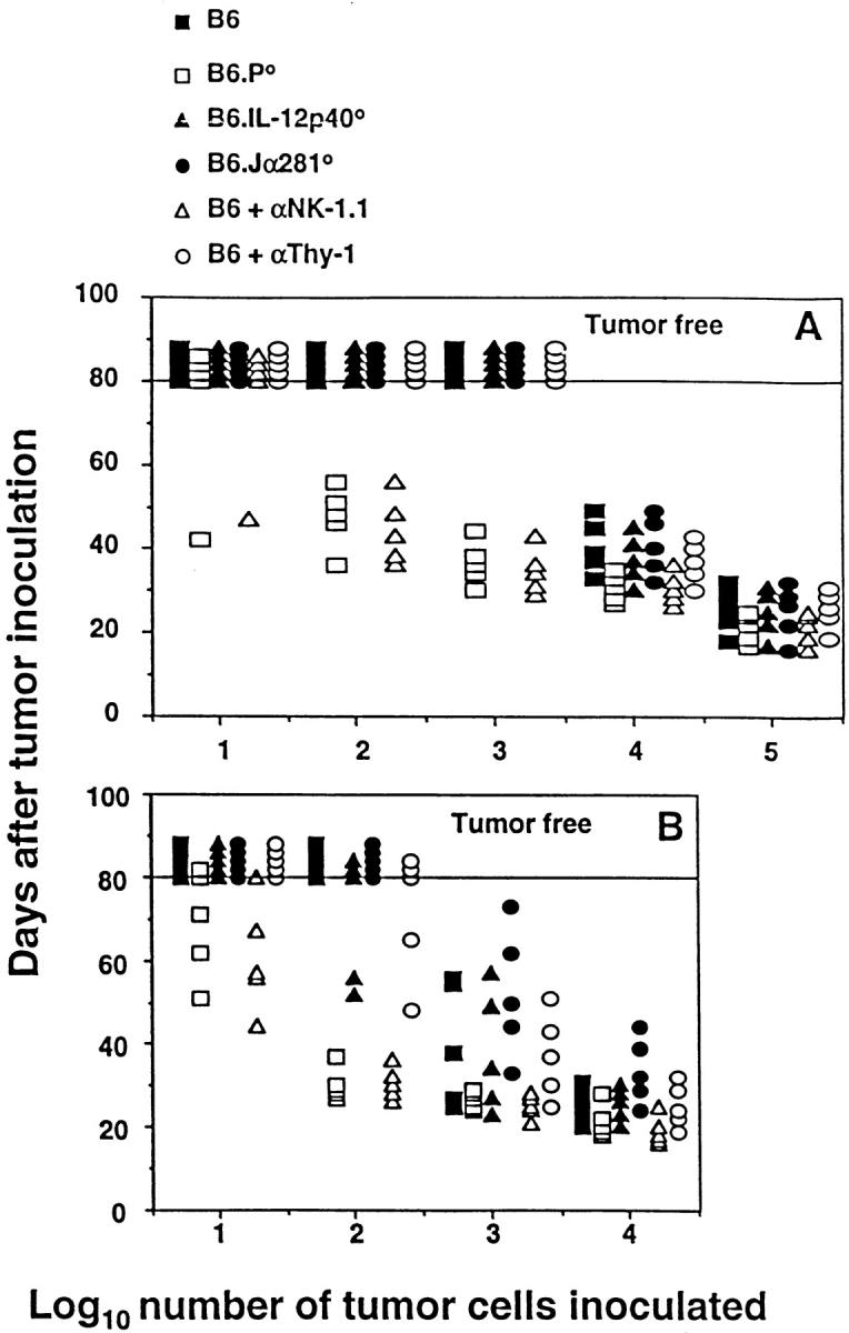
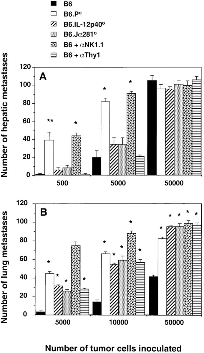
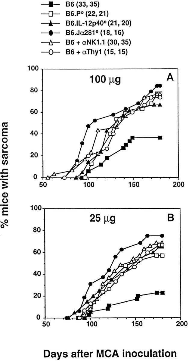
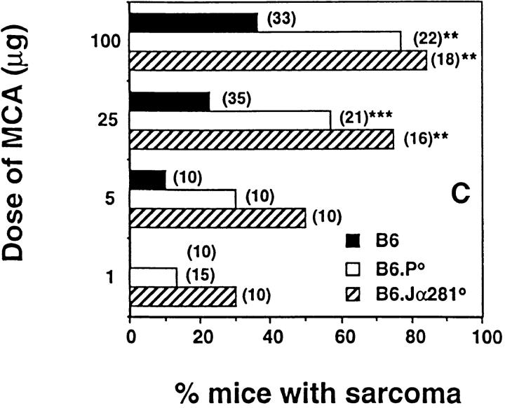
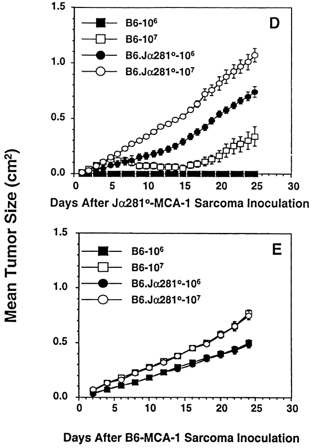
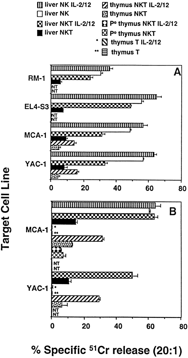
Natural tumor surveillance capabilities of the host were investigated in six different mouse tumor models where endogenous interleukin (IL)-12 does or does not dictate the efficiency of the innate immune response. Gene-targeted and lymphocyte subset-depleted mice were used to establish the relative importance of natural killer (NK) and NK1.1(+) T (NKT) cells in protection from tumor initiation and metastasis. In the models examined, CD3(-) NK cells were responsible for tumor rejection and protection from metastasis in models where control of major histocompatibility complex class I-deficient tumors was independent of IL-12. A protective role for NKT cells was only observed when tumor rejection required endogenous IL-12 activity. In particular, T cell receptor Jalpha281 gene-targeted mice confirmed a critical function for NKT cells in protection from spontaneous tumors initiated by the chemical carcinogen, methylcholanthrene. This is the first description of an antitumor function for NKT cells in the absence of exogenously administered potent stimulators such as IL-12 or alpha-galactosylceramide.


Figures

Figure 1
Figure 1
Elimination of intraperitoneally administered MHC class I–negative syngeneic tumors in vivo is mediated by NK cells and is IL-12 independent. B6, B6.P−/− (B6.P0), B6.IL-12p40−/− (B6.IL-12p400), or B6.Jα281−/− (B6.Jα2810) mice (five per group) were injected intraperitoneally with tumor cells (10–105) in 0.2 ml PBS, as indicated. Some groups of B6 mice were depleted of NK cells or T cells in vivo by treatment with mAb, 100 μg anti-NK1.1, or anti-Thy1, respectively, on days −2 and 0 (day of intraperitoneal tumor inoculation), and weekly thereafter. Mice were observed daily for tumor growth for 80 d by monitoring the development of ascites in mice. Individual mice are represented by each symbol. (A) EL4-S3 and (B) RM-1.
Figure 2
Figure 2
Innate control by NK and NKT cells in protection from tumor metastasis. (A) Groups of five B6, B6.P−/− (B6.P0), B6.IL-12p40−/− (B6.IL-12p400), or B6.Jα281−/− (B6.Jα2810) mice were injected intravenously with 500, 5,000, or up to 50,000 EL4-S3 tumor cells. 14 d later, their livers were removed and fixed in Bouin's solution, and surface lung metastases were counted with the aid of a dissecting microscope. Some groups of B6 mice were depleted of NK cells or T cells in vivo, as in the legend to Fig. 1. (B) Groups of five B6, B6.P−/− (B6.P0), B6.IL-12p40−/− (B6.IL-12p400), or B6.Jα281−/− (B6.Jα2810) mice were injected subcutaneously with RM-1 tumor cells (2 × 10⁶), and tumors were established for 9 d. At this time, subcutaneous tumors were surgically resected, and RM-1 cells were injected via the dorso-lateral tail vein. Mice were killed 14 d later, the lungs were removed and fixed in Bouin's solution, and surface lung metastases were counted with the aid of a dissecting microscope. Control experiments were performed by inoculating mice with RM-1 cells intravenously and counting lung metastases 14 d later. Some groups of B6 mice were depleted of NK cells or T cells in vivo, as in the legend to Fig. 1. The data for A and B were recorded as the mean (n = 5) number of metastases ± SEM. Significant differences from B6 group were determined by an unpaired t test to determine a two-tail P value (*P < 0.0001; **P < 0.005).
Figure 3
Figure 3
NKT cells protect mice from MCA-induced sarcoma. Groups of B6, B6.P−/− (B6.P0), B6.IL-12p40−/− (B6.IL-12p400), or B6.Jα281−/− (B6.Jα2810) mice (number of mice in parentheses) were injected subcutaneously in the hind flank with (A) 100 μg or (B) 25 μg MCA diluted in 0.1 ml corn oil. Mice were observed weekly for tumor development over the course of 50–180 d. Tumors >5 mm in diameter and demonstrating progressive growth over 3 wk were counted as positive. Some groups of B6 mice were depleted of NK cells or T cells in vivo, as in the legend to Fig. 1. In C, sarcoma development in B6, B6.P−/− (B6.P0), and B6.Jα281−/− (B6.Jα2810) mice was compared at several doses of MCA with data recorded at 180 d as a percentage of the mice in each group (in parentheses). Significant differences from the B6 group were determined by a Fisher's exact test (**P < 0.005; ***P < 0.02). Sarcomas derived from (D) B6.Jα281−/− mice (Jα2810-MCA-1) and (E) B6 mice (B6-MCA-1) were transplanted at 106 or 107 cells subcutaneously into groups of five B6.Jα281−/− or B6 mice. Tumor growth was measured daily with a caliper square as the product of two diameters. Results were recorded as the mean tumor size (in cm2) ± SEM, and are representative of two similar sarcomas transplanted into B6 and B6.Jα281−/− mice.
Figure 3
Figure 3
NKT cells protect mice from MCA-induced sarcoma. Groups of B6, B6.P−/− (B6.P0), B6.IL-12p40−/− (B6.IL-12p400), or B6.Jα281−/− (B6.Jα2810) mice (number of mice in parentheses) were injected subcutaneously in the hind flank with (A) 100 μg or (B) 25 μg MCA diluted in 0.1 ml corn oil. Mice were observed weekly for tumor development over the course of 50–180 d. Tumors >5 mm in diameter and demonstrating progressive growth over 3 wk were counted as positive. Some groups of B6 mice were depleted of NK cells or T cells in vivo, as in the legend to Fig. 1. In C, sarcoma development in B6, B6.P−/− (B6.P0), and B6.Jα281−/− (B6.Jα2810) mice was compared at several doses of MCA with data recorded at 180 d as a percentage of the mice in each group (in parentheses). Significant differences from the B6 group were determined by a Fisher's exact test (**P < 0.005; ***P < 0.02). Sarcomas derived from (D) B6.Jα281−/− mice (Jα2810-MCA-1) and (E) B6 mice (B6-MCA-1) were transplanted at 106 or 107 cells subcutaneously into groups of five B6.Jα281−/− or B6 mice. Tumor growth was measured daily with a caliper square as the product of two diameters. Results were recorded as the mean tumor size (in cm2) ± SEM, and are representative of two similar sarcomas transplanted into B6 and B6.Jα281−/− mice.
Figure 4
Figure 4
NKT cells directly lyse MCA-induced sarcoma. Thymus and liver NKT cells (NK1.1+TCR-β+) were sorted from B6 and B6.P−/− mice, and were examined in (A) 4-h and (B) 18-h 51Cr-release assays for direct lysis of a panel of labeled target cells as indicated. Some effector cells were cultured in IL-2 (50 U/ml) and IL-12 (20 pg/ml) throughout the course of the assay (+ IL-2/12). NKT effectors were tested at four different E/T ratios (20, 5, 1, 0.5), with an E/T ratio of 20:1 shown. In A, sorted liver NKT cells are compared with sorted liver NK cells. In B, sorted thymus NKT cells are with sorted thymus T cells (CD4+CD8 HSA). The data is recorded as the mean ± SEM, the spontaneous release of 51Cr was always <15% (subtracted from all tests), and each test was performed using duplicate samples. NT, not tested.

References

    1. Trinchieri G. Biology of natural killer cells. Adv. Immunol. 1989;47:187–376. - PMC - PubMed
    1. Talmadge J.E., Meyers K.M., Prieur D.J., Starkey J.R. Role of NK cells in tumour growth and metastasis in beige mice. Nature. 1980;284:622–624. - PubMed
    1. Karre K., Ljunggren H.G., Piontek G., Kiessling R. Selective rejection of H-2-deficient lymphoma variants suggests alternative immune defence strategy. Nature. 1986;319:675–678. - PubMed
    1. Salazar-Mather T.P., Ishikawa R., Biron C.A. NK cell trafficking and cytokine expression in splenic compartments after IFN induction and viral infection. J. Immunol. 1996;157:3054–3064. - PubMed
    1. Bendelac A., Rivera M.N., Park S.H., Roark J.H. Mouse CD1-specific NK1 T cellsdevelopment, specificity, and function. Annu. Rev. Immunol. 1997;15:535–562. - PubMed

Publication types

MeSH terms

Substances