. 2000 Mar 1;20(5):1657-65.
doi: 10.1523/JNEUROSCI.20-05-01657.2000.

Neurons regulate extracellular levels of amyloid beta-protein via proteolysis by insulin-degrading enzyme

Affiliations

Neurons regulate extracellular levels of amyloid beta-protein via proteolysis by insulin-degrading enzyme

K Vekrellis et al. J Neurosci. .

Abstract

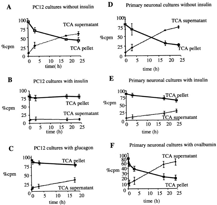
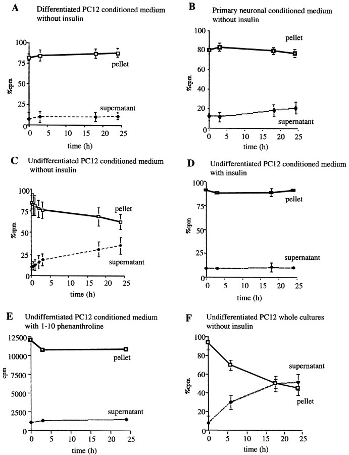
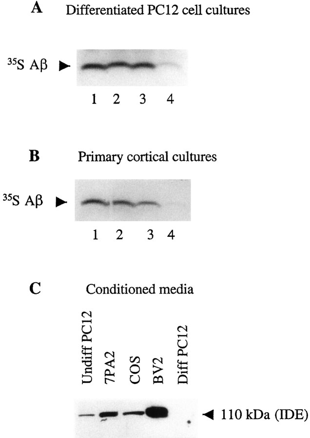
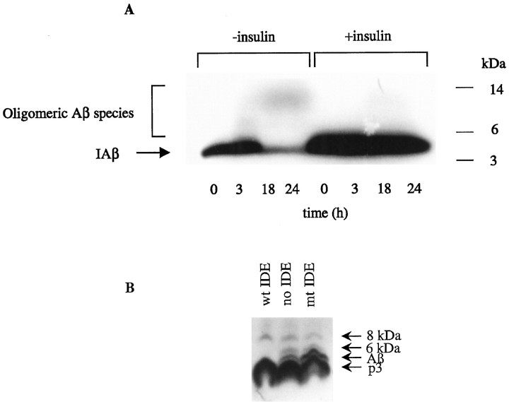
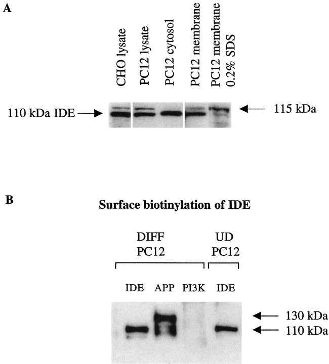
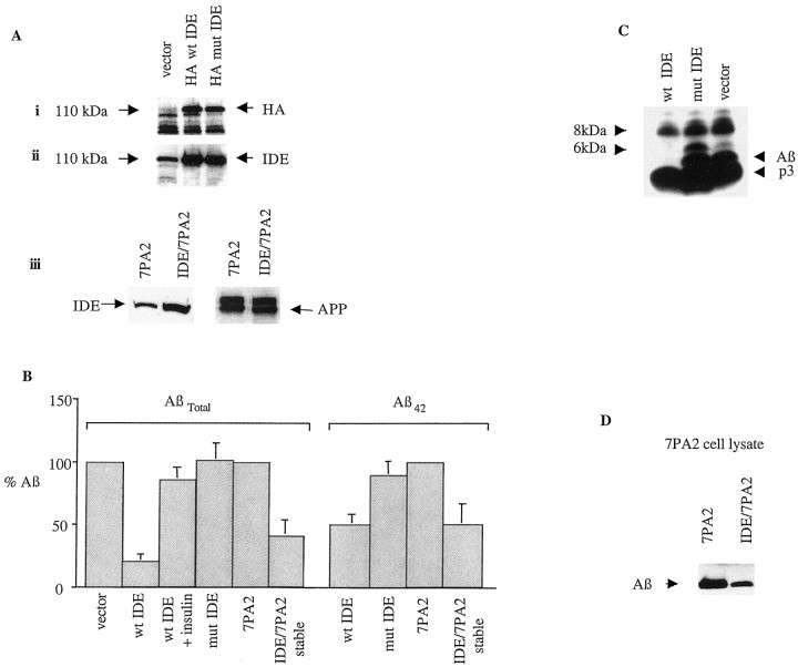
Progressive cerebral accumulation of amyloid beta-protein (Abeta) is an early and invariant feature of Alzheimer's disease. Little is known about how Abeta, after being secreted, is degraded and cleared from the extracellular space of the brain. Defective Abeta degradation could be a risk factor for the development of Alzheimer's disease in some subjects. We reported previously that microglial cells release substantial amounts of an Abeta-degrading protease that, after purification, is indistinguishable from insulin-degrading enzyme (IDE). Here we searched for and characterized a role for IDE in Abeta degradation by neurons, the principal cell type that produces Abeta. Whole cultures of differentiated pheochromocytoma (PC12) cells and primary rat cortical neurons actively degraded endogenously secreted Abeta via IDE. However, unlike that in microglia, IDE in differentiated neurons was not released but localized to the cell surface, as demonstrated by biotinylation. Undifferentiated PC12 cells released IDE into their medium, whereas after differentiation, IDE was cell associated but still degraded Abeta in the medium. Overexpression of IDE in mammalian cells markedly reduced the steady-state levels of extracellular Abeta(40) and Abeta(42), and the catalytic site mutation (E111Q) abolished this effect. We observed a novel membrane-associated form of IDE that is approximately 5 kDa larger than the known cytosolic form in a variety of cells, including differentiated PC12 cells. Our results support a principal role for membrane-associated and secreted IDE isoforms in the degradation and clearance of naturally secreted Abeta by neurons and microglia.


Figures

Fig. 1.
Fig. 1.
Degradation of Aβ in differentiated PC12 cell cultures and primary neurons is mediated by IDE. A–C, PC12 cells were differentiated in DMEM and 0.5% FBS supplemented with 100 ng/ml NGF for 5–7 d. Whole cultures were then incubated with 300 pm125I-Aβ in the absence (A) or presence of the known IDE substrates insulin (10 μm;B) or glucagon (10 μm;C). D–F, Primary mixed rat cortical cultures were maintained in DMEM supplemented with FBS and glucose for 3 weeks. For Aβ degradation assays, cultures were conditioned overnight and then incubated for up to 24 hr with 300 pm IAβ in the presence (D) or absence (E) of insulin or the presence of a control protein, ovalbumin (10 μm;F). Aliquots of CM were removed at the indicated times, and loss of intact peptide was assayed by TCA precipitation; the percents of total cpm recovered in supernatants and pellets are displayed. Curves represent means (± SEM) ofn = 4 experiments.
Fig. 2.
Fig. 2.
Differentiated neuronal cells do not release IDE into their medium. A, B, Differentiated PC12 cells (A) or 21 d primary mixed cortical cultures (B) were conditioned for 12 hr before the CM were collected and incubated for up to 24 hr with 300 pm IAβ.C–E, Undifferentiated PC12 culture medium that had been conditioned for 12 hr was similarly incubated with 300 pmIAβ in the absence (C) or presence (D) of 10 μm insulin or with 1 mm 1,10-phenanthroline (E).F, Whole cultures of undifferentiated PC12 cells were incubated with 300 pm IAβ in the absence of insulin. Aliquots of the CM were removed at the times indicated, and loss of intact IAβ was assayed by TCA precipitation. Curvesrepresent means (± SEM) of n = 4 experiments
Fig. 3.
Fig. 3.
Naturally secreted Aβ is degraded by a cell-associated form of IDE present in whole neuronal cultures but not in their conditioned media. A, Conditioned media of 7PA2 CHO cells labeled with [35S]Met (as a source of [35S]Aβ) were incubated at 37°C for 24 hr with either unconditioned 7PA2 medium (lane 1), unconditioned PC12 cell medium (lane 2), conditioned PC12 medium (lane 3), or differentiated PC12 whole cultures (lane 4). By the use of R1282 immunoprecipitation, labeled secreted Aβ could only be degraded by differentiated PC12 cell whole cultures and not by their conditioned medium alone. B, [35S]Met-labeled 7PA2 cell medium was incubated for 24 hr either with unconditioned 7PA2 medium (lane 1), mixed cortical cultures plus insulin (lane 2), conditioned medium of the cortical cultures (lane 3), or the whole cortical cultures (lane 4). Note that the starting amount of labeled Aβ just before the 24 hr incubation (time 0) is actually what remains after degradation in the 7PA2 cultures by IDE during the initial 16 hr conditioning period. We observed no further Aβ degradation after 16 hr of conditioning in 7PA2 cultures (data not shown). C,Thirty micrograms of NH4SO4-concentrated conditioned medium from each of the indicated cell types were separated by 10% Tris/Glycine SDS-PAGE and immunoblotted with the IDE-specific antibody IDE-1. Note the absence of IDE in the medium of differentiated PC12 cells. Diff, Differentiated.
Fig. 4.
Fig. 4.
IDE stimulates the oligomerization of iodinated synthetic Aβ but not naturally secreted Aβ in neuronal cultures.A, Primary mixed cortical cultures were conditioned in serum-free medium overnight and incubated with IAβ (300 pm) for up to 24 hr in the presence or absence of insulin (10 μm). Aliquots of the reaction mixtures were removed at the times indicated and characterized by 10–20% Tris/Tricine gel fluorography. Note that the loss of Aβ monomer and the formation of small amounts of SDS-stable higher molecular weight species are almost abolished by insulin. B, 7PA2 CHO cells were labeled with [35S]Met for 12 hr in the absence (no IDE) or presence of 100 ng of purified recombinant active IDE (wt IDE) or mutant (E111Q) IDE (mt IDE). CM were collected, immunoprecipitated with the Aβ antibody R1282, and assayed by SDS-PAGE and fluorography. Note that the 6 kDa species has been shown to be a modified form of Aβ monomer that migrates anomalously in SDS-PAGE gels; it always follows the behavior of the 4 kDa conventional Aβ monomer, in contrast to that of the 8 kDa Aβ dimer (Podlisny et al., 1998) (D. Walsh, R. Wong, and D. J. Selkoe, unpublished observations).
Fig. 5.
Fig. 5.
IDE can be detected on the surface of neuronal cells. A, Equal amounts of protein (30 μg) of CHO lysate as well as differentiated PC12 cytosol, lysate, membranes, and membranes extracted with 0.2% SDS were electrophoresed on 10% Tris/Glycine gels and Western blotted with the antibody IDE-1. Unlike cytosol in which only the 110 kDa IDE is detected, the whole lysate and membranes of PC12 cells also contain a higher (∼115 kDa) IDE-reactive band. Note that this higher band is also present in the CHO lysate.B, Differentiated and proliferating PC12 cells were biotinylated and lysed in STEN buffer, and equal amounts of protein were immunoprecipitated with the monoclonal antibody 9B12 against IDE (IDE), a monoclonal antibody specific to the cytosolic protein PI3-kinase (PI3K), or a polyclonal antibody (C7) to APP (APP). The immunoprecipitates were separated by SDS-PAGE and probed with neutravidin-HRP. Note that the large majority of biotinylated APP is the mature, N-plus O-glycosylated form (130 kDa), as expected. DIFF, Differentiated;UD, undifferentiated.
Fig. 6.
Fig. 6.
Expression of IDE markedly reduces steady-state levels of endogenous Aβ in the media. 7PA2 CHO cells were grown to 80% confluency before being transfected overnight with a pCMV expression vector encoding HA-tagged wild-type or mutant IDE or with vector alone. After transfection, the cells were refed with growth medium. In addition, 7PA2 CHO cells that were stably transfected with IDE were grown to 90% confluency and then refed with growth medium for 4 hr. A, Transient and stable transfection efficiency was quantitated by SDS-PAGE and autoradiography of the cell extracts using a specific monoclonal antibody to the HA epitope (i) as well as our polyclonal antibody IDE-1 (ii). APP background expression was quantitated using the specific polyclonal antibody C7 (iii). Note that endogenous IDE in the vector lane is solely detected by IDE-1, not anti-HA, as expected. B, Levels of endogenous Aβtotal (primarily Aβ40) and Aβ42 in the conditioned media of transiently and stably transfected cultures as indicated were established by ELISA.Verticalbars indicate means (± SEM). Values were normalized to vector alone or 7PA2 alone as 100%.C, 7PA2 cells were transiently transfected as described above and then starved in methionine-free medium for 2 hr before labeling with 100 μCi of [35S]me-thionine overnight. Conditioned media were immunoprecipitated with R1282 and assayed by SDS-PAGE autoradiography. In agreement with the ELISA results, wt IDE expression markedly reduced the levels of secreted monomeric Aβ. Note that the 6 kDa anomalously migrating monomeric Aβ isoform behaves like the 4 kDa monomer, in contrast to the 8 kDa dimer, which is primarily resistant to IDE. p3 is also resistant to IDE as reported previously (Qiu et al., 1998). D, Lysates from five 10 cm dishes of CHO cells stably transfected with APP or with both IDE and APP were precleared with APP antibodies C7 and B5, immunoprecipitated with R1280, and blotted with the monoclonal Aβ antibody 6E10. mut, Mutant.

References

    1. Akiyama H, Yokono K, Shii K, Ogawa W, Taniguchi H, Baba S, Kasuga M. Natural regulatory mechanisms of insulin degradation by insulin degrading enzyme. Biochem Biophys Res Commun. 1990;170:1325–1330. - PubMed
    1. Authier F, Bergeron JJ, Ou WJ, Rachubinski RA, Posner BI, Walton PA. Degradation of the cleaved leader peptide of thiolase by a peroxisomal proteinase. Proc Natl Acad Sci USA. 1995;92:3859–3863. - PMC - PubMed
    1. Authier F, Cameron PH, Taupin V. Association of insulin-degrading enzyme with a 70 kDa cytosolic protein in hepatoma cells. Biochem J. 1996a;319:149–158. - PMC - PubMed
    1. Authier F, Posner BI, Bergeron JJM. Insulin-degrading enzyme. Clin Invest Med. 1996b;19:149–160. - PubMed
    1. Baumeister H, Muller D, Rehbein M, Richter D. The rat insulin-degrading enzyme. Molecular cloning and characterization of tissue-specific transcripts. FEBS Lett. 1993;317:250–254. - PubMed

Publication types

MeSH terms