. 2000 Mar 1;20(5):1710-21.
doi: 10.1523/JNEUROSCI.20-05-01710.2000.

Reconstitution of muscarinic modulation of the KCNQ2/KCNQ3 K(+) channels that underlie the neuronal M current

Affiliations

Reconstitution of muscarinic modulation of the KCNQ2/KCNQ3 K(+) channels that underlie the neuronal M current

M S Shapiro et al. J Neurosci. .

Abstract

Channels from KCNQ2 and KCNQ3 genes have been suggested to underlie the neuronal M-type K(+) current. The M current is modulated by muscarinic agonists via G-proteins and an unidentified diffusible cytoplasmic messenger. Using whole-cell clamp, we studied tsA-201 cells in which cloned KCNQ2/KCNQ3 channels were coexpressed with M(1) muscarinic receptors. Heteromeric KCNQ2/KCNQ3 currents were modulated by the muscarinic agonist oxotremorine-M (oxo-M) in a manner having all of the characteristics of modulation of native M current in sympathetic neurons. Oxo-M also produced obvious intracellular Ca(2+) transients, observed by using indo-1 fluorescence. However, modulation of the current remained strong even when Ca(2+) signals were abolished by the combined use of strong intracellular Ca(2+) buffers, an inhibitor of IP(3) receptors, and thapsigargin to deplete Ca(2+) stores. Muscarinic modulation was not blocked by staurosporine, a broad-spectrum protein kinase inhibitor, arguing against involvement of protein kinases. The modulation was not associated with a shift in the voltage dependence of channel activation. Homomeric KCNQ2 and KCNQ3 channels also expressed well and were modulated individually by oxo-M, suggesting that the motifs for modulation are present on both channel subtypes. Homomeric KCNQ2 and KCNQ3 currents were blocked, respectively, at very low and at high concentrations of tetraethylammonium ion. Finally, when KCNQ2 subunits were overexpressed by intranuclear DNA injection in sympathetic neurons, total M current was fully modulated by the endogenous neuronal muscarinic signaling mechanism. Our data further rule out Ca(2+) as the diffusible messenger. The reconstitution of muscarinic modulation of the M current that uses cloned components should facilitate the elucidation of the muscarinic signaling mechanism.


Figures

Fig. 1.
Fig. 1.
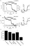
Inhibition of KCNQ2/KCNQ3 currents and generation of Ca2+ signals by a muscarinic agonist.A, Current amplitude at 0 mV in a tsA cell transfected with KCNQ2, KCNQ3, and M1 muscarinic receptors. Oxo-M (10 μm) and XE991 (50 μm) were bath-applied during the periods that are marked. The pipette solution contained 0.1 mm BAPTA. The inset shows the pulse protocol that was used and the current wave forms before and after the application of oxo-M. The dashed line in the current traces is the zero current level. Pulses were given every 4 sec.B, Microfluorometric measurement of [Ca2+]i in a tsA cell transfected with the M1 muscarinic receptor and bath-loaded with indo-1 as the AM ester for 20 min before measurements were taken. The cell was not patch-clamped and is different from that in A. Oxo-M (10 μm) was applied during the four periods that are marked.
Fig. 2.
Fig. 2.
Inhibition of KCNQ2/KCNQ3 currents by muscarinic agonists without Ca2+ signals. The holding current amplitude (filledcircles) is plotted for a tsA cell transfected with KCNQ2, KCNQ3, and M1 muscarinic receptors. The [Ca2+]i trace (bottom) was measured simultaneously from the fluorescence of indo-1 dialyzed into the cell from the patch pipette. Oxo-M (10 μm) was bath-applied during the periods that are shown. To prevent [Ca2+]i changes, the pipette solution contained the BACaPPS cocktail, which includes 20 mm BAPTA, 10 mm Ca2+, and 100 μg/ml PPS. Theinset shows the pulse protocol and current traces before and after the first application of oxo-M. The dashed line in the current traces is the zero current level. Pulses were given every 4 sec.
Fig. 3.
Fig. 3.
Depletion of internal stores by thapsigargin does not prevent the muscarinic inhibition of KCNQ2/KCNQ3 currents. Thapsigargin (2 μm) was bath-applied to cells transfected with KCNQ2, KCNQ3, and M1 muscarinic receptors to deplete internal Ca2+ stores, followed by the application of oxo-M (10 μm) to test for muscarinic inhibition.A, The pipette solution contained 0.1 mmBAPTA without PPS. Filledcircles are current amplitudes, and the line is the [Ca2+]i trace from indo-1 fluorescence. Thapsigargin, oxo-M, and XE991 were applied as shown by the horizontalbars. Selected current traces during the experiments are shown on the right. The dashed line in the current traces is the zero current level. B, The pipette contained the BACaPPS cocktail. Traces are as in A. C, Summary of KCNQ2/KCNQ3 channel inhibition under the various conditions as described in Results.
Fig. 4.
Fig. 4.
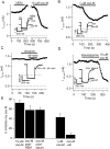
Muscarinic inhibition of KCNQ2/KCNQ3 currents is NEM-insensitive, uses M1 receptors, and is not mediated by protein kinases. Plotted in A–D are current amplitudes at the indicated holding potential in tsA cells transfected with KCNQ2, KCNQ3, and M1 receptors. The pipette solution contained the BACaPPS cocktail. A, NEM (50 μm) and oxo-M (10 μm) were bath-applied as indicated. The record contains a gap of 5 min. Current traces before NEM, after NEM, and after oxo-M applications are shown in the inset. Thedashed line in the current traces is the zero current level. B, Oxo-M (1 μm) was bath-applied as indicated. C, Atropine (100 μm) and oxo-M (1 μm) were bath-applied as indicated. D, Staurosporine (1 μm) and oxo-M (10 μm) were bath-applied as indicated. The pipette also contained 1 μm staurosporine. E, Summary of current inhibition under these conditions.
Fig. 5.
Fig. 5.
Voltage independence of muscarinic modulation.A, Families of current elicited by voltage steps from −70 to 40 mV, in 10 mV intervals, before and after the addition of 10 μm oxo-M to the bath. Cells were pretreated with staurosporine (1 μm) for 2 min before the addition of oxo-M. The holding potential and the tail current potential were −70 mV. The pipette solution contained the BACaPPS cocktail plus 1 μm staurosporine. The inhibition by oxo-M that is shown may be overestimated slightly because of run-down, although it was not excessive in this cell. Tail currents are marked byarrows. B, Amplitudes of the tail currents versus test potential for all experiments like those inA. Tail currents were quantified by measuring the average amplitude for 20 msec at 30 msec after repolarization to −70 mV. The data were fit with Boltzmann relations of the form: %I/Imax = 100 ·Imax/{1 + exp[(V1/2V)/k]}, whereV1/2 is the voltage that produces half-maximal activation of the conductance and k is the slope factor. Before oxo-M application, V1/2was −21 mV and k was 10.3 mV;Imax was set to unity. After oxo-M application, Imax was 0.26,V1/2 was −18 mV, and k was 10.3 mV.
Fig. 6.
Fig. 6.
TEA sensitivity of KCNQ2 and KCNQ3 homo- and heteromultimers. Shown are current amplitudes at −20 mV in cells transfected with KCNQ2 (A), KCNQ3 (B), or KCNQ2 and KCNQ3 (C). The pipette solution contained the BACaPPS cocktail. TEA was applied at the indicated concentrations. The dose–response data are summarized in D. The observations (symbols) were fit by a binding equation (curves) of the form: % Block = 100([TEA]/(K1/2 + [TEA])), whereK1/2 is the concentration for half-block. The fitted K1/2 was 174 μm for KCNQ2 and 224 mm for KCNQ3, but the data for KCNQ3 do not extend high enough for a good determination. For KCNQ2 plus KCNQ3, we first tried to fit the data as the sum of two Hill equations, withK1/2 values taken from the homomeric data (174 μm and 224 mm) (dottedline). We then fit the data by the sum of five binding equations for channels, with zero to four of each type of subunit (solid line). The distribution of channel arrangements was governed by the binomial distribution with a fit ratio of KCNQ2/KCNQ3 subunits of 0.35:0.65. The K1/2values for KCNQ2 and KCNQ3 were taken from the homomeric data, and the affinities for channels with the various subunit arrangements were calculated assuming energy additivity (see Results). The predictedK1/2 values for channels with one KCNQ2 and three KCNQ3 subunits, two KCNQ2 and two KCNQ3 subunits, and three KCNQ2 and one KCNQ3 subunits were 37.4, 6.24, and 1.04 mm, respectively. For all of the fits the maximal block was constrained to be 100%. The binomial modeling of the TEA data is qualitative and meant to demonstrate the expression of heteromeric versus homomeric channels. The data are not sufficient to determine the ratio of expressed KCNQ2 and KCNQ3 subunits with precision. Because theK1/2 value for TEA block of the KCNQ3 currents is not well determined, the heteromeric data also were fit by allowing that value to be a free parameter. This did not change the fit significantly.
Fig. 7.
Fig. 7.
KCNQ2 and KCNQ3 are modulated individually by oxo-M. A, Current amplitudes recorded with the BACaPPS cocktail at 0 mV in cells transfected with KCNQ2 and M1receptors. Oxo-M (10 μm) and XE991 (50 μm) were applied as indicated. B, A similar experiment but with cells transfected with KCNQ3 and M1 receptors.C, Current traces before and after the application of oxo-M. The dashed line shows the zero current level.D, Summary of inhibition with KCNQ2 or KCNQ3 as compared with results from KCNQ2/KCNQ3 channels taken from Figure3C. They are not significantly different at thep < 0.05 level.
Fig. 8.
Fig. 8.
KCNQ2 channels can be expressed in SCG neurons and modulated by the endogenous muscarinic pathway. Shown are amplitudes of the time-dependent current at −60 mV from voltage pulses given every 4 sec in SCG neurons. A, An uninjected neuron. TEA (1 mm) and oxo-M (10 μm) were bath-applied as indicated. Current traces are shown on the right in the control (a) and in the presence of TEA (b) or oxo-M (c). Thedashed line in the current traces is the zero current level. B, Similar experiment with a SCG neuron injected intranuclearly the previous day with plasmid for KCNQ2 (see Materials and Methods for a description of injections and a definition of current amplitude). C, Mean suppression of M current by TEA (black bars) and oxo-M (dashedbars). For uninjected and KCNQ2-injected cells the numbers of cells tested were 10 and 11, respectively. Two of the cells labeled uninjected were injected only with the GFP marker.

References

    1. Barhanin J, Lesage F, Guillemare E, Fink M, Lazdunski M, Romey G. KvLQT1 and IsK (minK) proteins associate to form the IKs cardiac potassium current. Nature. 1996;384:78–80. - PubMed
    1. Bean BP. Neurotransmitter inhibition of neuronal calcium currents by changes in channel voltage dependence. Nature. 1989;340:153–156. - PubMed
    1. Beech DJ, Bernheim L, Mathie A, Hille B. Intracellular Ca2+ buffers disrupt muscarinic suppression of Ca2+ current and M current in rat sympathetic neurons. Proc Natl Acad Sci USA. 1991;88:652–656. - PMC - PubMed
    1. Bernheim L, Beech DJ, Hille B. A diffusible second messenger mediates one of the pathways coupling receptors to calcium channels in rat sympathetic neurons. Neuron. 1991;6:859–867. - PubMed
    1. Bernheim L, Mathie A, Hille B. Characterization of muscarinic receptor subtypes inhibiting Ca2+ current and M current in rat sympathetic neurons. Proc Natl Acad Sci USA. 1992;89:9544–9548. - PMC - PubMed

Publication types

MeSH terms

Substances