. 2000 Mar 1;20(5):1931-40.
doi: 10.1523/JNEUROSCI.20-05-01931.2000.

Properties of horizontal and vertical inputs to pyramidal cells in the superficial layers of the cat visual cortex

Affiliations

Properties of horizontal and vertical inputs to pyramidal cells in the superficial layers of the cat visual cortex

Y Yoshimura et al. J Neurosci. .

Abstract

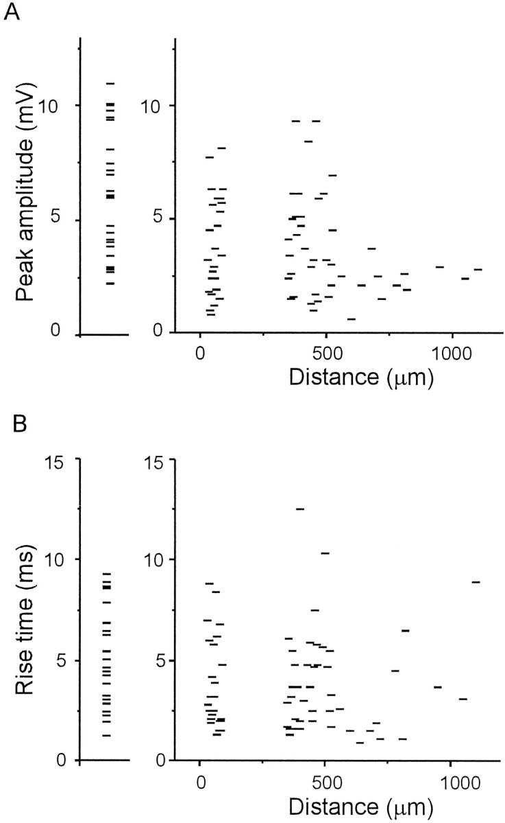
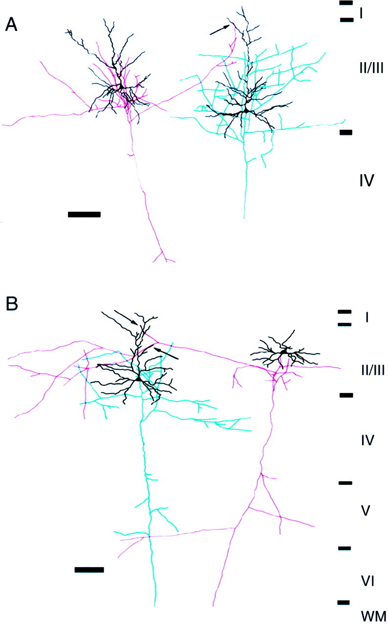
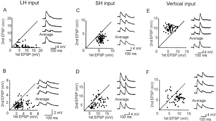
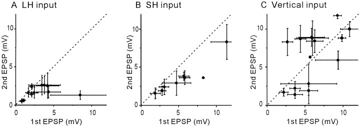
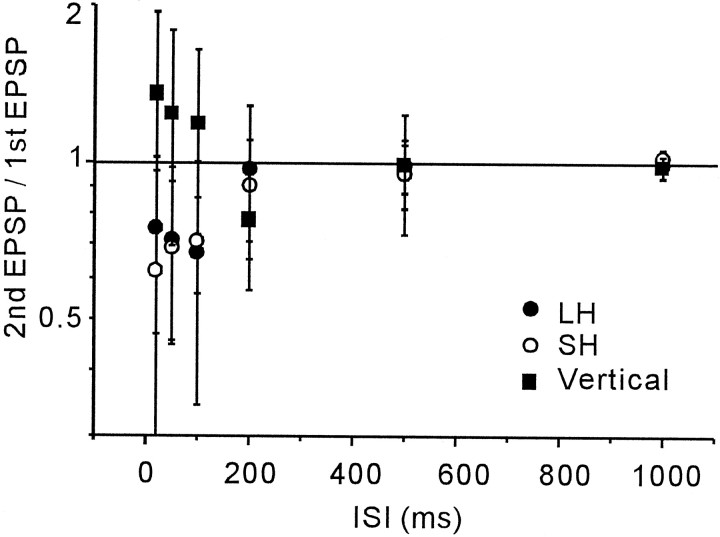
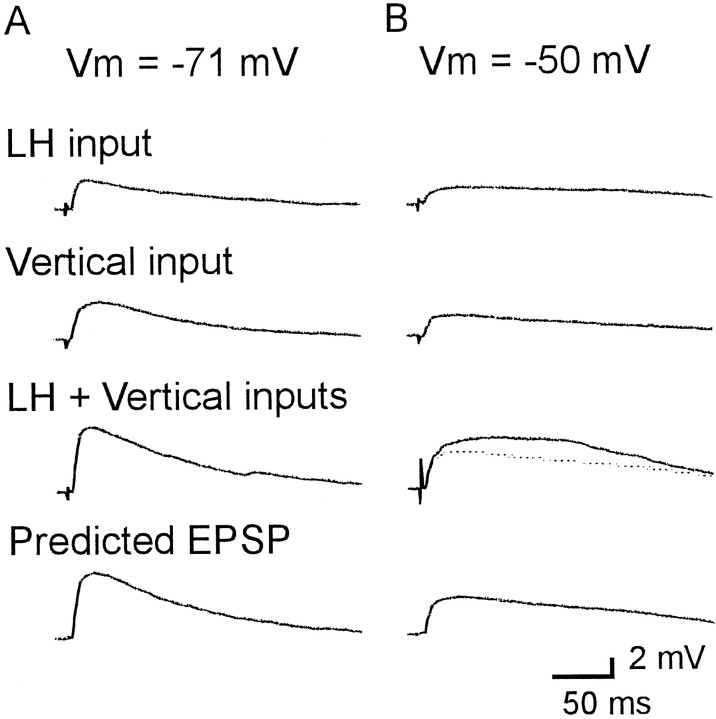
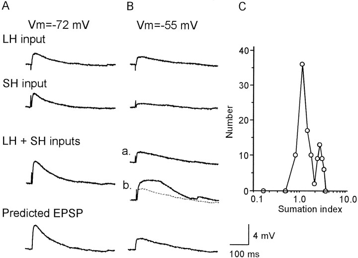
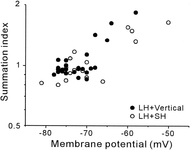
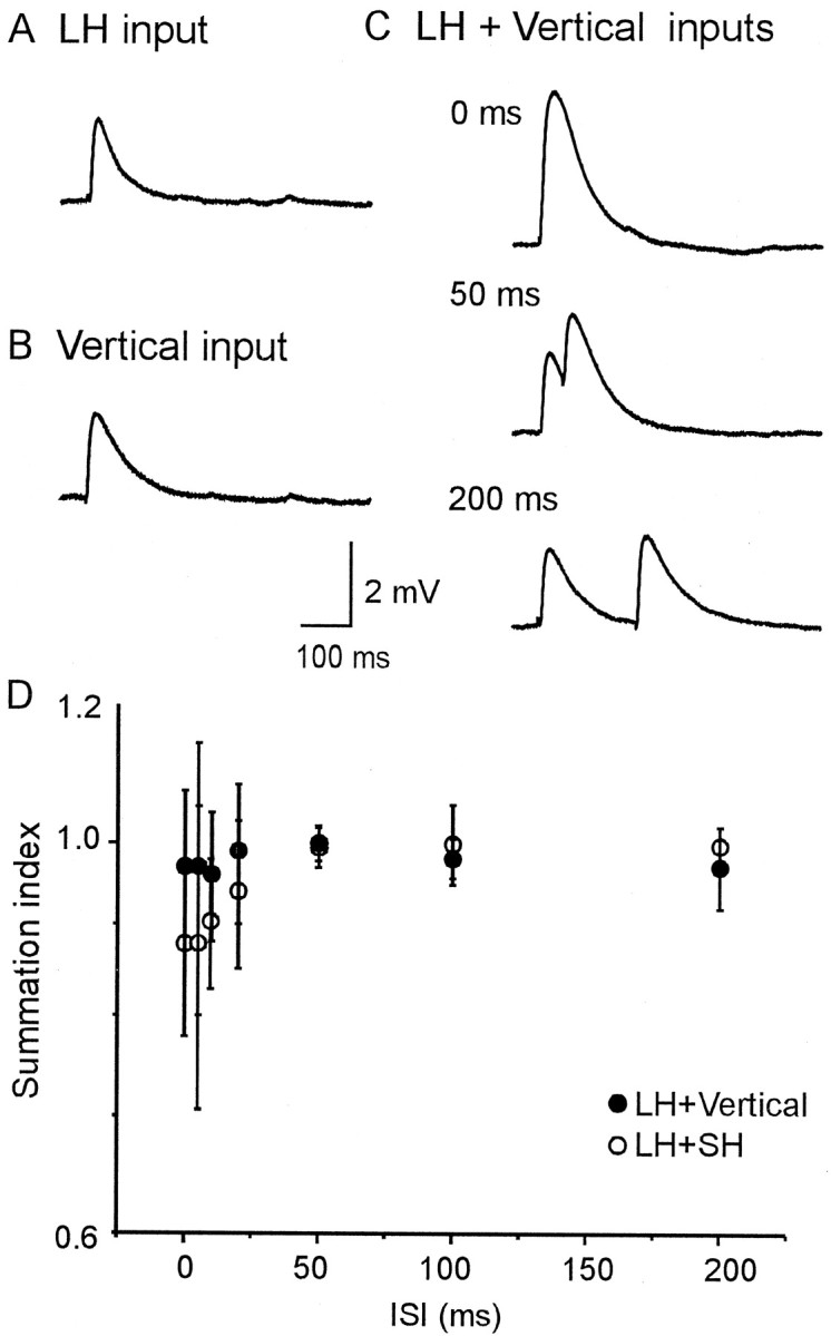
The purpose of this study is to elucidate the integrative input mechanisms of pyramidal cells receiving horizontally projecting axon collaterals (horizontal projection) and vertical input from layer IV. We performed whole-cell recordings from pyramidal cells in layer II/III and focally activated other single pyramidal cells monosynaptically connected via long-distance horizontal (LH) projections (the distance between presynaptic and postsynaptic cells was 350-1200 micrometer) in slice preparations of the kitten primary visual cortex. In addition, presynaptic single fibers in layer IV (vertical input) and/or short-distance horizontal (SH) inputs from neighboring single pyramidal cells (distance within 100 micrometer) in layer II/III were activated. Unitary EPSPs evoked by the activation of LH and SH connections had smaller amplitude and larger coefficient of variation than those evoked by stimulating the vertical input. Paired-pulse stimulation of the LH and SH inputs caused the depression of the second EPSP, whereas that of vertical inputs caused either facilitation or depression of the second EPSP. The EPSPs evoked by simultaneous activation of LH and vertical inputs summated linearly at the resting membrane potential. However, the EPSPs evoked by stimulation of the two inputs were nonlinearly (supralinearly) summated when the postsynaptic membrane was depolarized to a certain level. Similar EPSP interaction was observed in response to simultaneous activation of the LH and SH inputs.


Figures

Fig. 1.
Fig. 1.
Schematic arrangement of electrodes (A) and examples of unitary EPSPs evoked by focal stimulation of horizontal (C) and vertical (D) input pathways. When cell a ord in A was stimulated, clear EPSPs were recorded from a pyramidal cell in the superficial layers, as shown inC. No EPSP was evoked by the stimulation of cellb and c in A. The distance between the stimulated and recorded cells was 60, 350, 410, and 440 μm for cells a–d, respectively. B, The stimulus–response curve of EPSPs obtained from the cell shown inC and D. Each dotindicates the mean peak amplitude of EPSPs evoked by the stimulation of the cell d. Error bars indicate SDs. The abrupt increase of the EPSP amplitude at the stimulus intensity of 13 V suggests that the EPSPs were unitary. D, EPSPs evoked by minimal stimulation of the layer IV.
Fig. 2.
Fig. 2.
Relationship between the distance separating the presynaptic and postsynaptic cells in layer II/III and the electrophysiological parameters of unitary EPSPs. In the graphs on theright, the peak amplitude (A) and rise time (B) of the unitary EPSPs are plotted as a function of the distance between the recorded and stimulated cells. Each horizontal tick represents data for each pair of horizontally connected cells. The connections between cells separated by distances of <100 μm and 350–1200 μm were defined as SH and LH connections, respectively. Because of sampling bias, cell pairs separated by the distance of 100–350 μm are absent. Note the lack of correlation between the distance and the amplitude or rise time of the EPSPs. The horizontal ticks in the graphs on theleft represent the mean peak amplitude (A) and rise time (B) of the EPSPs evoked by vertical input stimulation.
Fig. 3.
Fig. 3.
Reconstruction of a pair of connected pyramidal cells in the superficial layers of the kitten visual cortex.A, B, Two examples of pairs connected via long-distance horizontal axon collaterals. The soma and dendrites are shown in black. The axons of the presynaptic and postsynaptic cells are shown in red andblue, respectively. Boutons of axon collaterals of the presynaptic cell are closely apposed to the apical dendrites (arrows) of the postsynaptic cell. Electrophysiological data of the pairs in A and B are shown in Figures 7 and 10A–C, respectively. Scale bars, 100 μm.
Fig. 4.
Fig. 4.
Effects of paired-pulse stimulation in six experiments. Successive stimulations at an ISI of 100 msec were applied to LH (A, B), SH (C, D), and vertical (E, F) input pathways. Each graph illustrates the data from one pair of cells. Diagonal lines indicate that the amplitudes of the first EPSPs are the same as those of the second. Insets show three individual EPSPs and the average (20–30 trials).
Fig. 5.
Fig. 5.
A summary of paired-pulse experiments on the three different input pathways. Each dot denotes the average value from one experiment. Horizontal andvertical bars show the SD of the amplitudes of the first and second EPSPs, respectively. Dashed diagonal linesindicate that the amplitudes of the first EPSPs are the same as those of the second. The second EPSPs were significantly depressed compared to the first in 9 of 11 cell pairs for paired-pulse stimulation of LH input and in all eight pairs for that of SH input. Paired-pulse stimulation of vertical input resulted in either significant facilitation (6 of 15) or depression (7 of 15) of the second EPSPs.
Fig. 6.
Fig. 6.
ISI dependence of paired-pulse depression and facilitation. The mean ratios (± SD) of the amplitudes of the second EPSPs to those of the first EPSPs are plotted against the tested ISIs for LH (n = 7), SH (n = 6), and vertical (n = 6) connections. Paired-pulse stimulations with ISIs of <100 msec, on average, induced second EPSP depressions for LH inputs and SH inputs and facilitation for vertical inputs.
Fig. 7.
Fig. 7.
Interaction between EPSPs elicited by activation of LH and vertical inputs. A, Recordings at the resting membrane potential (−71 mV). B, Recordings under the depolarized condition (−50 mV). Single stimulation applied to LH inputs (top traces) or vertical inputs (second traces) induced unitary EPSPs. EPSPs were recorded after simultaneous stimulation of LH and vertical inputs (third traces). Predicted EPSPs (bottom traces) were calculated as a simple mathematical sum of the top two traces. EPSPs evoked by concurrent activation of the two inputs was found to be a linear sum of the two individual EPSPs at the resting membrane potential (A). However, supralinear summation was observed under the condition of a depolarized membrane (B). The EPSP induced by simultaneous stimulation of the two inputs (thick line) and the predicted EPSP (dotted line) were superimposed in the third trace in B. The morphology of this cell pair is shown in Figure 3A.
Fig. 8.
Fig. 8.
Interaction between unitary EPSPs evoked by activation of LH (720 μm) and SH (60 μm) inputs. The data shown in A–C were obtained from one cell.A, EPSPs evoked by simultaneous activation of the two inputs were found to be linearly summated at the resting membrane potential (−72 mV). B, Linear (a) and supralinear (b) summations were observed under the condition of a depolarized membrane (−55 mV). InBb, the recorded EPSP (thick line) and the predicted EPSP (dotted line) were superimposed.C, The distribution of the summation index calculated from the EPSPs recorded at −55 mV; data represent those from the experiment shown in B. Note the two clear peaks, the ones on the left and right correspond to linear and nonlinear summation, respectively. Other conventions are the same as in Figure 7.
Fig. 9.
Fig. 9.
Induction of nonlinear interaction by concurrent activation of two inputs depends on the depolarization of membrane potential. Summation indices of cells were plotted against the membrane potential of postsynaptic cells. When two inputs are summated linearly, the SI takes the value of 1. Each point represents data from one cell (n = 26 for LH and vertical input interaction,n = 16 for LH and SH input interaction). For the interaction between LH and SH inputs, linear (or slightly sublinear) summation was observed at the resting membrane potential. Induction of supralinear summation is clearly dependent on the depolarization of the postsynaptic cell for both input interactions.
Fig. 10.
Fig. 10.
Temporal interaction between LH and vertical or SH inputs. EPSPs evoked by individual stimulation (A, B) and concurrent stimulation of the two inputs (C).C, EPSPs were linearly summated by concurrent stimulation of both LH and vertical inputs with all tested ISIs.Vm = −77 mV. The morphology of this pair is shown in Figure 3B. D, The mean SIs (± SD) were plotted against the tested ISIs. The graph shows pooled data from six cells for LH and vertical inputs and six cells for LH and SH inputs. The SIs of the interaction between LH and SH inputs are <1 for ISIs of <50 msec.

References

    1. Abbott LF, Varela JA, Sen K, Nelson SB. Synaptic depression and cortical gain control. Science. 1997;275:220–224. - PubMed
    1. Akasaki T, Yoshimura Y, Sato H. Modulation of visual response properties by stimulus outside of the classical receptive field in cat area 17. Neurosci Res. 1998;22:S180.344.
    1. Allen C, Stevens CF. An evaluation of causes for unreliability of synaptic transmission. Proc Natl Acad Sci USA. 1994;91:10380–10383. - PMC - PubMed
    1. Blakemore C, Tobin EA. Lateral inhibition between orientation detectors in the cat's visual cortex. Exp Brain Res. 1972;15:439–440. - PubMed
    1. Callaway EM, Katz LC. Emergence and refinement of clustered horizontal connections in cat striate cortex. J Neurosci. 1990;10:1134–1153. - PMC - PubMed

Publication types

LinkOut - more resources