. 2000 Mar 1;20(5):1952-63.
doi: 10.1523/JNEUROSCI.20-05-01952.2000.

Dual serotonin (5-HT) projections to the nucleus accumbens core and shell: relation of the 5-HT transporter to amphetamine-induced neurotoxicity

Affiliations

Dual serotonin (5-HT) projections to the nucleus accumbens core and shell: relation of the 5-HT transporter to amphetamine-induced neurotoxicity

P Brown et al. J Neurosci. .

Abstract

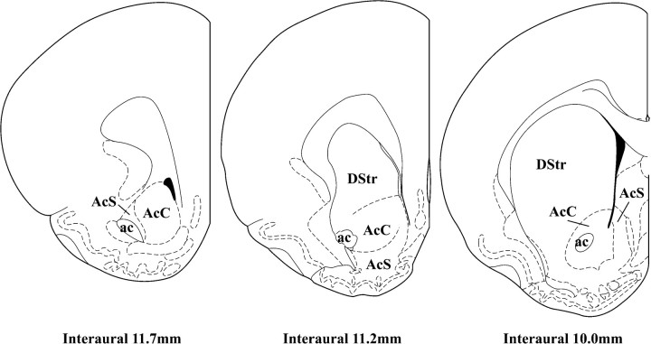
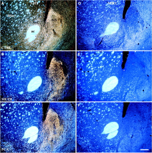
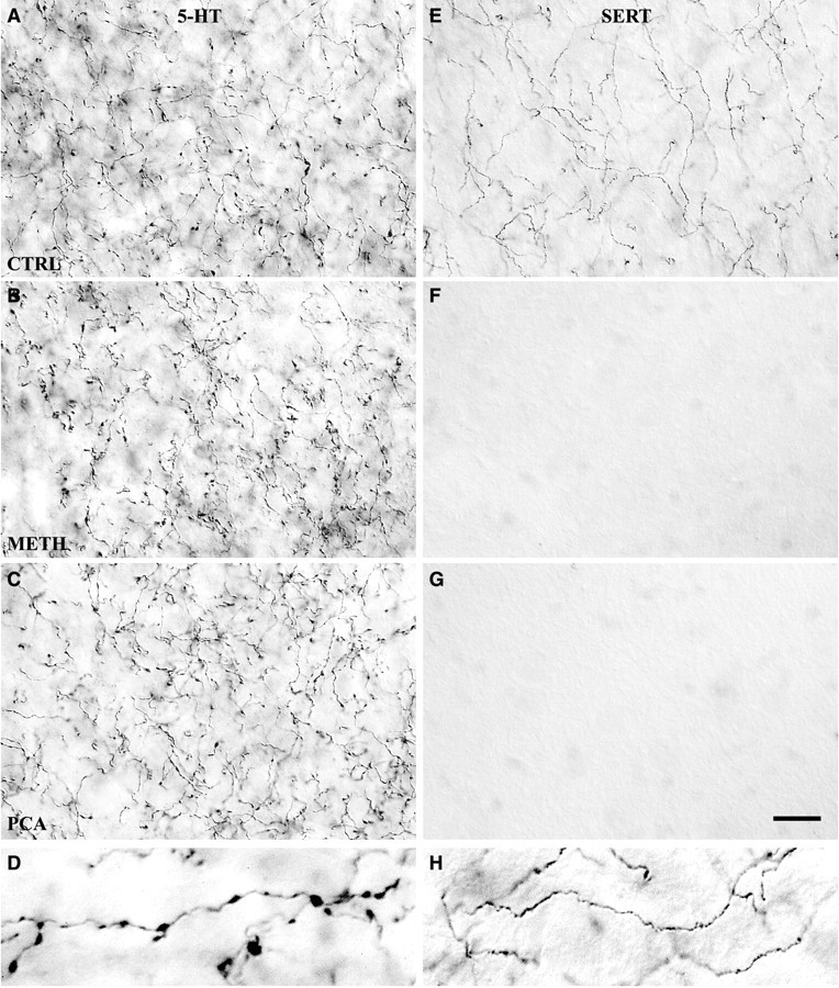
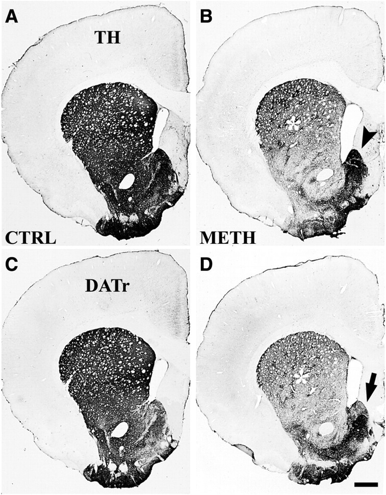
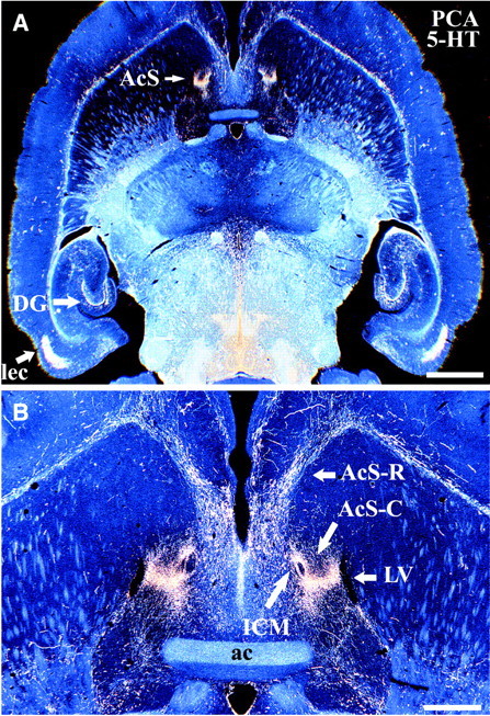
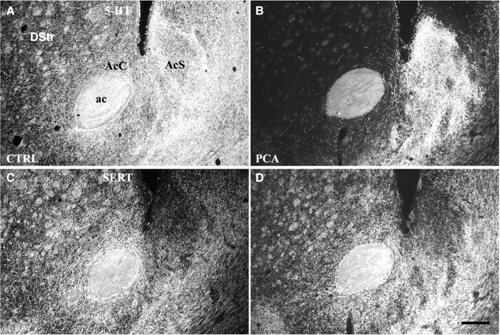
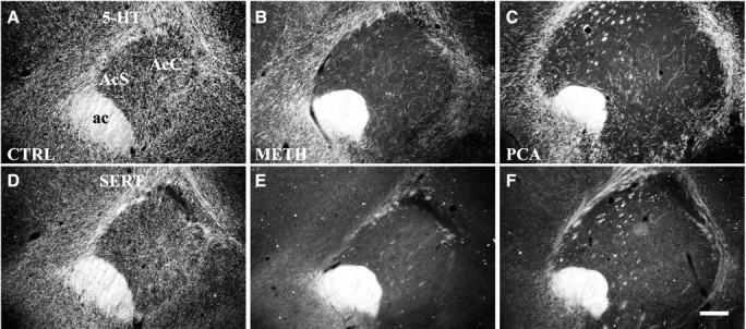
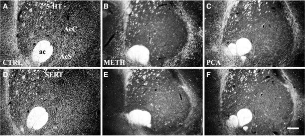
Dopamine release in the nucleus accumbens (NAc) has been implicated as mediating the rewarding effects of stimulant drugs; however, recent studies suggest that 5-HT release may also contribute. In an effort to assess the role of 5-HT in drug-mediated reward, this study analyzed the serotonergic innervation of NAc using immunocytochemistry for 5-HT and the 5-HT transporter (SERT). We report that in control rats the NAc receives two distinct types of 5-HT axons that differ in regional distribution, morphology, and SERT expression. Most regions of the NAc are innervated by thin 5-HT axons that express SERT, but in the caudal NAc shell nearly all 5-HT axons lack SERT and have large spherical varicosities. Two weeks after methamphetamine or p-chloroamphetamine (PCA) treatment, most 5-HT axons in dorsal striatum and NAc have degenerated; however, the varicose axons in the shell appear intact. These drug-resistant 5-HT axons that lack SERT densely innervate the caudal one-third of the accumbens shell, the same location where dopamine axons are spared after methamphetamine. Moreover, 4 hr after PCA, the varicose axons in the caudal shell retain prominent stores of 5-HT, whereas 5-HT axons in the rest of the NAc are depleted of neurotransmitter. The results demonstrate that two functionally different 5-HT projections innervate separate regions of the NAc and that selective vulnerability to amphetamines may result from differential expression of SERT. We postulate that action potentials conducted from the raphe nuclei can release 5-HT throughout the NAc, whereas transporter-mediated release induced by stimulant drugs is more restricted and unlikely to occur in the caudal NAc shell.


Figures

Fig. 1.
Fig. 1.
Drawings of three coronal levels through nucleus accumbens and dorsal striatum. Line drawings show the rostral pole (interaural 11.7 mm), intermediate division (interaural 11.2 mm), and caudal (interaural 10.0 mm) division of the nucleus accumbens, where the serotonergic innervation was analyzed in this study (from Paxinos and Watson, 1998). ac, Anterior commissure;AcC, nucleus accumbens core; AcS, nucleus accumbens shell; DStr, dorsal striatum.
Fig. 2.
Fig. 2.
Differential vulnerability of 5-HT axons in NAc shell and core. Dark-field images (coronal) show the effects of Meth (4 × 20 mg/kg) or PCA (2 × 10 mg/kg) on 5-HT-IR (A–C) and SERT-IR (D–F) axon terminals in the caudal division of the nucleus accumbens (interaural 10.0 mm). In control animals the 5-HT axons in NAc core and dorsal striatum express SERT, whereas most axons in the caudal shell are SERT-negative. After treatment with either drug, 5-HT axons in the core and dorsal striatum are ablated, whereas those in the shell are spared, demonstrating that SERT-negative axons are selectively resistant to amphetamine neurotoxicity. Drug-treated animals were killed 14 d after treatment, and sections were processed for immunocytochemistry. A, D, Saline-treated; B, E, Meth-treated; C, F, PCA-treated. Scale bar, 300 μm. ac, Anterior commissure; AcC, nucleus accumbens core; AcS, nucleus accumbens shell;DStr, dorsal striatum. (Note: “dorsal striatum” in the rat refers to the caudate–putamen complex as distinct from ventral striatum.)
Fig. 3.
Fig. 3.
Two morphological types of 5-HT axons in caudal NAc shell differ in SERT expression and in response to Meth and PCA. High-magnification images show the effect of Meth (4 × 20 mg/kg) or PCA (2 × 10 mg/kg) on 5-HT-IR (A–C) and SERT-IR (E–G) axon terminals in the caudal shell of the nucleus accumbens. Most 5-HT axons in the NAc shell have large varicosities (D) and lack SERT expression; these axons are unaffected by either drug (A–C). In control rats, a smaller number of thin axons in the shell lack varicosities and express SERT (E,H); the SERT-IR axons are ablated by either drug (F, G). Drug-treated animals were killed 14 d after drug treatment, and sections were processed by ICC to demonstrate 5-HT or SERT expression. Images were photographed with a 100× oil immersion objective using DIC. Scale bar, 15 μm. Enlarged images in thebottom row (at twice the magnification) show examples of varicose 5-HT-IR axon terminals (D) or thin nonvaricose SERT-IR axon terminals (H).A, E, Saline-treated; B, F, Meth-treated;C, G, PCA-treated.
Fig. 4.
Fig. 4.
Vulnerability of dopamine axons in striatum and nucleus accumbens to methamphetamine toxicity. Bright-field images show the effect of Meth (4 × 20 mg/kg) on TH-IR and DATr-IR axon terminals in dorsal striatum and nucleus accumbens. After Meth treatment, TH-IR is considerably reduced in midstriatum and in the NAc core (B), reflecting DA axonal degeneration (asterisk), whereas in the NAc shell most DA axons are spared (arrowhead). DATr-IR axons (D) showed a similar pattern of axonal degeneration. The location of spared DA axons at this level of the NAc shell (arrow) matches that of drug-resistant 5-HT axons (see Fig. 2B,C). Drug-treated animals were killed 14 d after treatment, and striatal sections were processed for immunohistochemistry. A, C, Saline-treated control;B, D, Meth-treated. Scale bar, 700 μm.
Fig. 5.
Fig. 5.
PCA-resistant 5-HT axons have a highly restricted localization in the caudal NAc shell. Low-magnification dark-field images show the distribution of drug-resistant varicose 5-HT-IR (SERT-negative) axon terminals in rat brain after PCA (2 × 10 mg/kg) treatment. Note the dense, restricted innervation by spared 5-HT-IR axons in the caudal NAc shell, as in dentate gyrus and entorhinal cortex (A). Higher magnification (B) reveals that the spared 5-HT axons in the caudal shell are situated between the lateral ventricle (laterally) and the island of Calleja magna (medially). Animals were killed 14 d after PCA treatment, and horizontal sections at the level of the NAc shell were processed for 5-HT immunocytochemistry. Scale bars:A, 2.0 mm; B, 600 μm.ac, Anterior commissure; AcS-C, caudal nucleus accumbens shell; AcS-R, rostral nucleus accumbens shell; DG, dentate gyrus; ICM, island of Calleja magna; lec, lateral entorhinal cortex;LV, lateral ventricle.
Fig. 6.
Fig. 6.
PCA acutely depletes 5-HT from serotonergic axons in NAc core but not in the caudal shell. Dark-field images (coronal) demonstrate PCA-induced (1 × 10 mg/kg) 5-HT depletion from axon terminals in the core of nucleus accumbens, whereas axons in the caudal shell remain intensely immunoreactive for 5-HT (B). Although the 5-HT-IR axons in the core are depleted, unaltered SERT-IR staining (D) demonstrates that these axons have not degenerated at this survival time. Animals were killed 4 hr after PCA treatment, and sections were processed for 5-HT and SERT ICC. A, B, 5-HT-IR axon terminals; C, D, SERT-IR axon terminals. Scale bar, 300 μm. ac, Anterior commissure; AcC, nucleus accumbens core; AcS, nucleus accumbens shell;DStr, dorsal striatum. A, C, Saline-treated; B, D, PCA-treated.
Fig. 7.
Fig. 7.
5-HT innervation of the rostral pole of the nucleus accumbens. Neurotoxic effects of Meth (4 × 20 mg/kg) or PCA (2 × 10 mg/kg) on 5-HT-IR (A–C) and SERT-IR (D–F) axon terminals in the rostral pole of the NAc (interaural 11.7 mm) are shown in dark-field images. Most 5-HT axons in the rostral pole are lost after treatment. A small number of 5-HT-IR axons are spared, and these are SERT-negative (B-F). Animals were killed 14 d after drug treatment, and adjacent sections were processed for 5-HT and SERT immunocytochemistry. A, D, Saline-treated; B, E, Meth-treated; C, F, PCA-treated. Scale bar, 300 μm. ac, anterior commissure; AcC, nucleus accumbens core; AcS, nucleus accumbens shell.
Fig. 8.
Fig. 8.
5-HT innervation of the intermediate division of the nucleus accumbens. Neurotoxic effects of Meth (4 × 20 mg/kg) or PCA (2 × 10 mg/kg) on 5-HT-IR (A–C) and SERT-IR (D–F) axon terminals in the intermediate division of the nucleus accumbens are shown in dark-field images (interaural 11.2 mm). Thin, SERT-positive serotonergic axons predominate at this intermediate level of NAc (A, D). As in the rostral pole, most 5-HT axons in the intermediate division are lost after drug treatment. A small number of 5-HT axons are spared, and these are SERT-negative (B–F). Animals were killed 14 d after drug treatment, and adjacent sections were processed for 5-HT and SERT immunocytochemistry. A, D, Saline-treated; B, E, Meth-treated; C, F, PCA-treated. Scale bar, 300 μm. ac, anterior commissure; AcC, nucleus accumbens core;AcS, nucleus accumbens shell.

References

    1. Axt KJ, Molliver ME. Immunocytochemical evidence for methamphetamine-induced serotonergic axon loss in the rat brain. Synapse. 1991;9:302–313. - PubMed
    1. Axt KJ, Molliver ME, Qian Y, Blakely RD. Subtypes of 5-HT axons differ in their expression of serotonin transporter. Soc Neurosci Abstr. 1995;21:865.
    1. Benloucif S, Galloway MP. Facilitation of dopamine release in vivo by serotonin agonists: studies with microdialysis. Eur J Pharmacol. 1991;200:1–8. - PubMed
    1. Berridge CW, Stratford TL, Foote SL, Kelley AE. Distribution of dopamine-β-hydroxylase-like immunoreactive fibers within the shell subregion of the nucleus accumbens. Synapse. 1997;27:230–241. - PubMed
    1. Blue ME, Yagaloff KA, Mamounas LA, Hartig PR, Molliver ME. Correspondence between 5-HT2 receptors and serotonergic axons in rat neocortex. Brain Res. 1988;453:315–328. - PubMed

Publication types

MeSH terms