. 2000 Mar 1;20(5):1990-2002.
doi: 10.1523/JNEUROSCI.20-05-01990.2000.

A proprioceptive role for an exteroceptive mechanoafferent neuron in Aplysia

Affiliations

A proprioceptive role for an exteroceptive mechanoafferent neuron in Aplysia

D Borovikov et al. J Neurosci. .

Abstract

Afferent regulation of centrally generated activity is likely to be more complex than has been established. We show that a neuron that is an exteroceptor can also function as a proprioceptor. We study the Aplysia neuron B21. Previous data suggest that B21 functions as an exteroceptor during the radula closing/retraction phase of ingestive feeding. We show that the tissue innervated by B21, the subradula tissue (SRT), is innervated by a motor neuron (B66) and that B66-induced SRT contractions trigger centripetal spikes in B21. Thus, B21 is also a proprioceptor. To determine whether exteroceptive and proprioceptive activities occur during the same phase of ingestive feeding, we further characterize B66. We show that B66 stimulation does not close or retract the radula. Instead it opens it. Moreover, B66 is electrically coupled to other opening/protraction neurons. Finally, we elicit motor programs in semi-intact preparations and show that during radula opening/protraction we observe B66 activity, SRT contractions, and spikes in B21 that can be eliminated if B66 is indirectly hyperpolarized. B21 is, therefore, likely to act as an exteroceptor during one phase of ingestive feeding and as a proprioceptor during the antagonistic phase. Previous experiments have shown that centripetal spikes in B21 are only transmitted to one follower if they are "gated in" by depolarization. During ingestive programs B21 is centrally depolarized during closing/retraction, but it is not depolarized during opening/protraction. We sought to determine whether there are other followers that receive B21 input when it is not centrally depolarized. We found one such cell. Moreover, we found that stimulation of B21 during radula opening/protraction significantly decreases the duration of this phase of behavior. Thus, proprioceptive activity in B21 is likely to have an impact on motor programs.


Figures
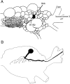
Fig. 1.
Fig. 1.
Morphology of B66. A, Schematic drawing of the caudal surface of the desheathed buccal ganglion. The position of the filledcell indicates the position of B66. Neurons B1/2 and B6 are labeled as reference cells. The drawing is after Hurwitz et al. (1997). CBC,Cerebral–buccal connective. B, Drawing of B66 injected with carboxyfluorescein dye. Note that B66 has an axon in buccal nerve 3.
Fig. 2.
Fig. 2.
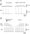
The B66 and SRT neuromuscular junction. A, Left, Excitatory junctional potentials recorded intracellularly from SRT fibers (top) induced by intracellular stimulation of B66 (bottom) in normal ASW.Right, Same experiment with the buccal ganglion bathed in a 3 × Mg+2 and 3 × Ca+2 solution. B, Inhibition of SRT contractions by hexamethonium. SRT contractions were measured with an isotonic force transducer (top) and elicited by intracellular stimulation of B66 (bottom).Left, Application of hexamethonium.Right, Washout with ASW.
Fig. 3.
Fig. 3.
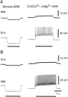
Neurons B66 and B61/62 are electrically coupled.A, Left, In normal ASW hyperpolarizing current injected into B61/62 (horizontalbarunderbottomtrace) resulted in a hyperpolarization of B66 (toptrace). Right, When depolarizing current was injected into B61/62 in a low-calcium solution, which blocks synaptic transmission, depolarization and coupling potentials were still apparent in B66. B, The same manipulations with current injected into B66 are shown. (This experiment was performed with two electrodes in the neuron that was hyperpolarized or depolarized and one electrode in the “follower” neuron.).
Fig. 4.
Fig. 4.
Neurons B66 and B48 are electrically coupled.A, Left, Hyperpolarizing current injected into B48 (horizontalbarunderbottomtrace) resulted in a hyperpolarization of B66 (toptrace).Right, When depolarizing current was injected into B48 in a low-calcium solution, depolarization and coupling potentials were still apparent in B66. B, The same manipulations with current injected into B66 are shown.
Fig. 5.
Fig. 5.
Neurons B66 and B13 are electrically coupled.A, Left, Hyperpolarizing current injected into B13 (horizontalbarunderbottomtrace) resulted in a hyperpolarization of B66 (toptrace).Right, When depolarizing current was injected into B13 in a low-calcium solution, depolarization and coupling potentials were also still apparent in B66. B, The same manipulations with current injected into B66 are shown.
Fig. 6.
Fig. 6.
Radula movements observed in a semi-intact preparation as a result of B66 stimulation. Left, Before B66 stimulation. Right, After stimulation. Note that stimulation of B66 results in an ipsilateral movement in which one-half of the radula moves away from the midline.
Fig. 7.
Fig. 7.
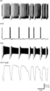
Contractions of the SRT occur during the opening/protraction phase of ingestive motor programs. A motor program was elicited by applying carbachol (10−3m) to the cerebral ganglion, which was in an isolated subchamber. The topthree traces are intracellular recordings from B8 (a motor neuron that produces radula closing), B4/5 (a multifunction neuron active during the radula closing/retraction phase of behavior), and B66. Thebottomtrace shows contractions of the SRT measured with an isotonic force transducer. Contractions of the SRT and activity in B66 were out-of-phase with activity in B4/5 and high-frequency activity in B8.
Fig. 8.
Fig. 8.
Hyperpolarization of B66 changes the size of SRT contractions during ingestive motor programs. Injecting depolarizing current into the command-like neuron CBI-2 induced a motor program. Thebottomthreetraces are intracellular recordings from CBI-2, B4/5, and B66. Thetoptrace shows contractions of the SRT measured with an isotonic force transducer. During one cycle of the program B66 was hyperpolarized. In four out of five preparations contractions of the SRT were not observed while B66 was hyperpolarized. In one preparation contractions of the SRT were reduced in size but not completely abolished. When hyperpolarization of B66 was released, SRT contractions returned.
Fig. 9.
Fig. 9.
Stimulation of B66 elicits contractions of the SRT and centripetal spikes in B21. A, Left, B66 was stimulated (horizontalbarundermiddletrace), and corresponding contractions of the SRT were monitored with an isotonic force transducer (bottomtrace). Activity in the sensory neuron B21 (toptrace) was observed. Right, This activity persisted when the buccal ganglion was placed in a low-calcium solution, indicating that B21 responses can be recorded in the absence of central synaptic activity.B, Left, B21 at resting potential is shown.Right, B21 was hyperpolarized. Note that when B21 was hyperpolarized spikes became smaller, as would be expected if they were centripetally generated.
Fig. 10.
Fig. 10.
Centripetal spikes in B21 occur as the SRT contracts, unless tension is developed slowly. SRT contractions were elicited when B66 was stimulated so that it generated two spikes or six spikes. (Action potentials in B66 are not shown.) Left, Two spikes in B66 generated an SRT contraction in which tension developed slowly. Under these conditions centripetal spikes were not triggered in B21. Right, In contrast, six spikes in B66 generated a contraction with a faster contraction rate, and action potentials were observed in B21 during the time that the SRT was contracting. Dashedlines facilitate the alignment of spikes in B21 and the SRT contraction. Leftand right are from the same preparation.
Fig. 11.
Fig. 11.
Action potentials are observed in neuron B21 during the opening/protraction phase of ingestive motor programs that are likely to be centripetally generated. An ingestive motor program was elicited by applying carbachol to the cerebral ganglion. Thetoptrace is an intracellular recording from B21, and the middletrace is a recording from B61/62 (a motor neuron that produces radula opening/protraction that is electrically coupled to B66). Thebottomtrace is an intracellular recording from the radula closing/retraction interneuron B64. Note that action potentials were observed in B21 while B61/62 was active [i.e., during the opening/protraction (O/P) phase of the motor program] and while B64 was active [i.e., during the closing/retraction (C/R) phase of the motor program]. During one cycle of the program B61/62 was hyperpolarized (horizontalbar). Because B61/62 and B66 are electrically coupled, hyperpolarization of B61/62 hyperpolarizes B66. Under these conditions spikes were not observed in B21 during opening/protraction, suggesting that these spikes were generated peripherally as a result of the contraction of the SRT.
Fig. 12.
Fig. 12.
Schematic diagram illustrating contacts between a normal B21 and B8 and between B21 and B64. Note that B21 is a bipolar or pseudobipolar neuron with a medial process that branches. One branch of the medial process crosses the midline and terminates in the contralateral buccal ganglion; the second branch projects to the periphery (including the SRT). B21's lateral process terminates in the ipsilateral hemiganglion in the vicinity of B8 and B64. Previous data suggest that B21 makes contact with B8 via its lateral process as shown. B21 could make contact with B64 via its lateral process (1), or via a more medial connection (2).
Fig. 13.
Fig. 13.
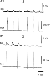
Effect of lesioning the lateral process of B21 on PSPs in B8 (B) and coupling potentials in B64 (A). Spikes in B21 were elicited by brief depolarization of its soma. A1, B1,Traces before the lateral process was lesioned are shown. A2, B2, Electrodes were removed from neurons, and the lateral process of B21 was lesioned. Cells were then reimpaled, and responses to stimulation of B21 were recorded. Note that coupling potentials in B64 are not significantly changed in size whereas PSPs in B8 are abolished. A and Bare from different preparations. B21 was at its resting potential (approximately −60 mV) in both experiments. Both experiments were performed in 3 × Ca2+ ASW so that PSPs would be more visible.
Fig. 14.
Fig. 14.
Verification of lesions. Carboxyfluorescein dye was injected into the left and right B21 neurons in a single buccal ganglion at the conclusion of a physiological experiment (see Fig. 13). The lateral process of theleft B21 was severed, whereas the rightB21 remained intact. The normal morphology of B21 can be seen by looking at the right cell. Note its lateral process (L), its soma (S), and its medial process (M). That the lesion was effective in this experiment can be seen by comparing the lateral processes of the right versus the leftB21. RN, Radula nerve. Scale bar, 500 μm.
Fig. 15.
Fig. 15.
Centripetal spikes are attenuated in the soma and lateral process of B21 when it is at its resting membrane potential. A1, Centripetal spikes were triggered when B21 was at its resting membrane potential (approximately −60 mV).Left, Resulting responses were recorded from the medial process of B21 (i.e., ∼200 μm from the soma). Right, Responses were also simultaneously recorded from the soma of B21. Note that responses recorded from the soma are smaller in amplitude.A2, Centripetal spikes were triggered when B21 was depolarized by ∼15 mV. Left, Responses recorded from the medial process are shown. Right, Responses recorded from the soma are shown. Note that responses are now similar in amplitude. Recordings shown in A1 and A2were made from the same preparation. The dottedlines facilitate amplitude comparisons.B1, Centripetal spikes were triggered when B21 was at its resting membrane potential (approximately −60 mV). Responses were simultaneously recorded from the soma (left) and the lateral process (i.e., ∼175 μm from the soma;right). Note that responses recorded from the lateral process are smaller. B2, Centripetal spikes were triggered when B21 was depolarized by ∼15 mV. Left, A response from the soma is shown. Right, A response from the lateral process is shown. Note that both spikes are increased in amplitude. Recordings shown in B1 and B2were made from the same preparation. Recordings in A andB are from two different preparations.
Fig. 16.
Fig. 16.
Central depolarization increases the size of B21-induced PSPs in B8 but less dramatically alters the size of B21-induced coupling potentials in B64. A, Spikes in B21 were elicited by brief depolarization of its soma. Left, B21 was at its resting potential (i.e., approximately −60 mV).Right, B21 was depolarized by 20 mV. B, Spikes in B21 were elicited by mechanical stimulation of the SRT.Left, B21 was at its resting potential.Right, B21 was depolarized by 20 mV.
Fig. 17.
Fig. 17.
Activity in B21 during the radula opening/protraction phase of an ingestive motor program can affect temporal characteristics of the motor program. Ingestive activity was induced when CBI-2 was intracellularly stimulated with brief pulses in bursts (toprow oftraces). CBI-2's intraburst firing frequency was 8 Hz, and bursts were elicited approximately once a minute. That widespread activity was indeed triggered in the feeding circuitry can be seen from the intracellular recording from B21 (secondrow of traces from thetop), from the intracellular recording from B64 (thirdrow of traces from the top), from the extracellular recording from the I2 nerve (fourthrow oftraces from the top), and from the extracellular recording from the radula nerve (RN;bottomrow of traces). The duration of the opening/protraction phase of the motor program was monitored via the extracellular recording from the I2 nerve, which contains the processes of the opening protraction interneurons and motor neurons B61/62 and B31/32. The closing/retraction phase of the motor program is most clearly marked by the activity in B64. As is apparent in AC this type of CBI-2 stimulation triggers one cycle of a two-phase motor program.A, Control (i.e., current was not injected into B21) traces are shown. B21 receives depolarizing input during closing/retraction but does not spike. B, B21 was stimulated with brief current pulses at 20 Hz after the initiation of opening/protraction. B21 stimulation was terminated shortly after the retraction phase was initiated. Note that the duration of the opening/protraction phase of the motor program was reduced.C, Ingestive activity was again triggered without spiking in B21. The duration of opening/protraction returned to its control value. Calibration: vertical, 40 mV;horizontal, 5 sec.

References

    1. Alexeeva A, Borovikov D, Miller MW, Rosen SC, Cropper EC. Effect of a serotonergic extrinsic modulatory neuron (MCC) on radula mechanoafferent function in Aplysia. J Neurophysiol. 1998;80:1609–1622. - PubMed
    1. Baxter DA, Patel VC, Susswein AJ, Bryne J. Computational model of a multifunctional central pattern generator (CPG) that underlies consummatory feeding behavior in Aplysia. Soc Neurosci Abstr. 1997;23:1044.
    1. Borovikov D, Rosen SC, Cropper EC. SCP-containing sensory neurons in Aplysia are peripherally activated as a result of motor neuron activity. Soc Neurosci Abstr. 1997;23:1045.
    1. Busches A, El Manira A. Sensory pathways and their modulation in the control of locomotion. Curr Opin Neurobiol. 1998;18:733–739. - PubMed
    1. Cattaert D, Clarac F. Presynaptic inhibition in crayfish primary afferents. In: Rudomin P, Romo R, Mendell LM, editors. Presynaptic inhibition and neural control. Oxford UP; New York: 1998. pp. 192–205.

Publication types