. 2000 Mar 15;28(6):1313-21.
doi: 10.1093/nar/28.6.1313.

Interaction of the yeast DExH-box RNA helicase prp22p with the 3' splice site during the second step of nuclear pre-mRNA splicing

Affiliations

Interaction of the yeast DExH-box RNA helicase prp22p with the 3' splice site during the second step of nuclear pre-mRNA splicing

D S McPheeters et al. Nucleic Acids Res. .

Abstract

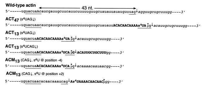
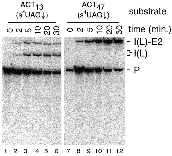
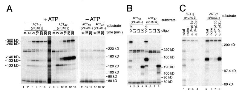
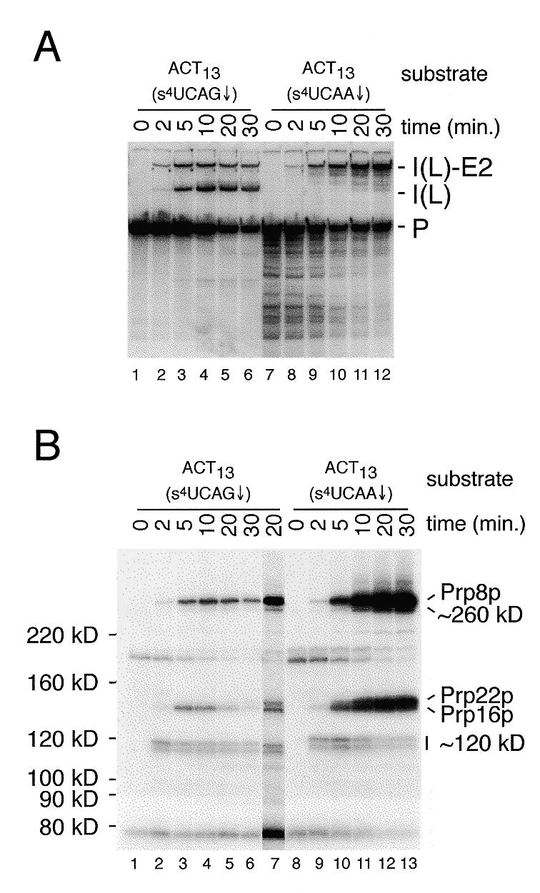
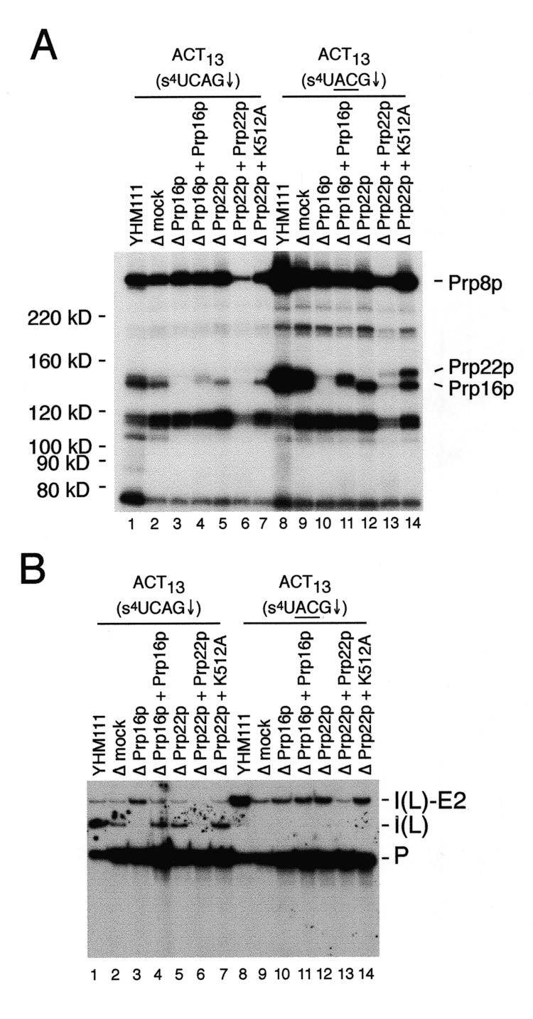
Using site-specific incorporation of the photo-chemical cross-linking reagent 4-thiouridine, we demonstrate the previously unknown association of two proteins with yeast 3' splice sites. One of these is an unidentified approximately 122 kDa protein that cross-links to 3' splice sites during formation of the pre--spliceosome. The other factor is the DExH-box RNA helicase, Prp22p. With substrates functional in the second step of splicing, only very weak cross-linking of Prp22p to intron sequences at the 3' splice site is observed. In contrast, substrates blocked at the second step exhibit strong cross-linking of Prp22 to intron sequences at the 3' splice site, but not to adjacent exon sequences. In vitro reconstitution experiments also show that the association of Prp22p with intron sequences at the 3' splice site is dependent on Prp16p and does not persist when release of mature mRNA from the spliceosome is blocked. Taken together, these results suggest that the 3' splice site of yeast introns is contacted much earlier than previously envisioned by a protein of approximately 120 kDa, and that a transient association of Prp22p with the 3' splice site occurs between the first and second catalytic steps.


Figures

Figure 1
Figure 1
Sequences of the branch site-3′ splice site region of the yeast actin pre-mRNA derivatives. The sequence of the region encompassing the branch site/3′ splice site region of the wild-type yeast actin pre-mRNA is shown for reference with the branch site adenosine in upper case. For each pre-mRNA, the branch site and 3′ splice site consensus sequences are underlined, the position of 3′ splice site cleavage is indicated by a downward arrow, and the 3′ exon sequence is shown in italics. Spacing between the branch sequence and 3′ splice site is measured from the branch site adenosine to the position of 3′ splice site cleavage. For each pre-mRNA constructed by three- or four-way in vitro RNA ligations, the sequence of the 5′ and 3′ RNA fragments are shown in lower case, the sequence of the unmodified, or s4U containing, oligo RNAs are shown in bold-face upper case and the position of the single 32P label is indicated by an asterisk. The ACT13(s4UCAA↓) and ACT13(s4UACG↓) substrates are identical to the ACT13(s4UCAG↓) substrate depicted here, except for a one or two base substitution in the 3′ splice site. The 3′ splice site mutant ACM13 (ACG↓) substrates are identical to the wild-type ACM13(CAG↓) substrates shown here, except for a two base substitution in the 3′ splice site.
Figure 2
Figure 2
Splicing activity of actin pre-mRNA derivatives containing s4U in the 3′ splice site sequence. The ACT13(s4UAG↓) and ACT47(s4UAG↓) substrates were incubated in splicing extract for the indicated times in the presence of 2 mM ATP. RNA species are labeled as follows: P, pre-mRNA; I(L)-E2, intron lariat-exon2 intermediate; I(L), intron lariat product.
Figure 3
Figure 3
(A) Analysis of proteins that cross-link to the 3′ splice sites of the ACT13(s4UAG↓) and ACT47(s4UAG↓) substrates. Aliquots of the splicing reactions shown in Figure 2 were subjected to cross-linking and the resulting 32P-labeled proteins analyzed on a 6% SDS–polyacrylamide gel. The positions of molecular weight markers are shown on the right and the approximate apparent molecular weights of the major ATP-dependent cross-linked proteins (see text) indicated on the left. Lane 7 shows a longer exposure of lane 5 to clearly illustrate the weak cross-linking of the ~140 kDa species to the ACT13(s4UAG↓) substrate. (B) Analysis of 3′ splice site cross-linking using splicing extracts depleted of the U1, U2 or U6 snRNAs. Extracts were pretreated with the indicated oligonucleotides, followed by incubation with the indicated substrates for 10 min, cross-linking and analysis on a 6% SDS–polyacrylamide gel. (C) Immunoprecipitation analysis of proteins that cross-link to the 3′ splice site. Samples from either 5 min splicing reactions done with the ACT13(s4UAG↓) substrate or a 20 min splicing reaction done with the ACT47(s4UAG↓) substrate were used for immunoprecipitation following cross-linking. For each substrate, a portion of each reaction was removed for visualization of the total cross-linked proteins (lanes 1 and 5) prior to immunoprecipitation with either anti-Prp16p or Prp22p anti-sera. In lanes 2 and 6 anti-sera was omitted to detect non-specific binding of 32P-labeled, cross-linked proteins to the protein A–agarose alone. A 6% SDS–polyacrylamide gel was used.
Figure 4
Figure 4
Splicing activity and analysis of proteins that cross-link to a wild-type and a 3′ splice site mutant pre-mRNA substrate. (A) Splicing assays were performed for the times indicated with either the wild-type ACT13(s4UCAG↓) substrate (lanes 1–6) or the 3′ splice site mutant ACT13(s4UCAA↓) substrate (lanes 7–12) in the presence of 2 mM ATP. RNA species are labeled as in Figure 2. (B) Aliquots of each of the splicing reactions shown in (A) were cross-linked and the resulting 32P-labeled proteins analyzed on a 6% SDS–polyacrylamide gel. Lane 7 is from a darker exposure of the 20 min time point with the ACT13(s4UCAG↓) substrate. On the original autoradiograph, clear separation of Prp16p and Prp22p is visible. In the comparison of proteins cross-linked to the ACT13(s4UCAG↓) and ACT13(s4UCAA↓) substrates, note that the relative migration of some of the cross-linked species seen with the 3′ splice site mutant is decreased due to the increased size (18 versus 24 nt) of the 32P-labeled RNase T1 fragment.
Figure 5
Figure 5
Cross-linking analysis in depleted extracts. (A) Cross-linking assays were performed in the indicated control (YHM111), mock depleted (Δmock), Prp16p (ΔPrp16p) or Prp22p depleted (ΔPrp22p), or reconstituted extracts using the indicated substrates. All samples were analyzed following a 10 min incubation at 23°C. Cross-linked 32P-labeled proteins in control and reconstituted extracts were analyzed on a 6% SDS–polyacrylamide gel. In this experiment, an undiluted, undialyzed sample of recombinant His-tagged Prp22p protein was used that caused an overall inhibition of splicing activity as well as cross-linking. In other experiments (data not shown), normal splicing activity and cross-linking to the HIS-tagged Prp22p at levels comparable to the K512A mutant protein are observed using 5- or 10-fold dilutions of the recombinant protein. On the original autoradiograph, clear separation of the His-tagged Prp16p and Prp22p in the tightly spaced doublet in lane 11 is visible. (B) Splicing assays of the samples used for cross-link analysis. RNA species are labeled as in Figure 2.
Figure 6
Figure 6
Cross-linking of proteins to the wild-type and mutant ACM13 substrates containing s4U at position –4 within the intron (lanes 1 and 2) or position +2 within the 3′ exon (lanes 3 and 4). Cross-linking assays were performed after incubation of reaction in the presence of 2 mM ATP for 12 min and samples were analyzed on a 7% SDS–polyacrylamide gel.

References

    1. Staley J.P. and Guthrie,C. (1998) Cell, 92, 315–326. - PubMed
    1. Moore M.J., Query,C.C. and Sharp,P.A. (1993) In Gesteland,R. and Atkins,J. (eds), The RNA World. Cold Spring Harbor Laboratory Press, Cold Spring Harbor, NY, pp. 303–358.
    1. Umen J.G. and Guthrie,C. (1995) RNA, 1, 869–885. - PMC - PubMed
    1. Spingola M., Grate,L., Haussler,D. and Ares,M.,Jr (1999) RNA, 5, 221–234. - PMC - PubMed
    1. Luukkonen B.G. and Seraphin,B. (1997) EMBO J., 16, 779–792. - PMC - PubMed

Publication types

MeSH terms