. 2000 Mar 15;28(6):1340-7.
doi: 10.1093/nar/28.6.1340.

Effects of RNA secondary structure on cellular antisense activity

Affiliations

Effects of RNA secondary structure on cellular antisense activity

T A Vickers et al. Nucleic Acids Res. .

Abstract

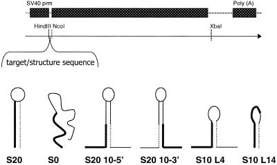
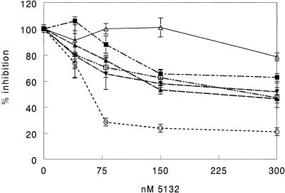
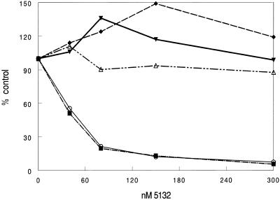
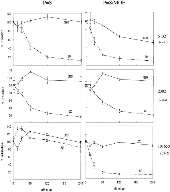
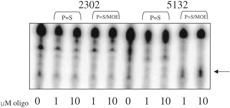
The secondary and tertiary structures of a mRNA are known to effect hybridization efficiency and potency of antisense oligonucleotides in vitro. Additional factors including oligonucleotide stability and cellular uptake are also thought to contribute to antisense potency in vivo. Each of these factors can be affected by the sequence of the oligonucleotide. Although mRNA structure is presumed to be a critical determinant of antisense activity in cells, to date little direct experimental evidence has addressed the significance of structure. In order to determine the importance of mRNA structure on antisense activity, oligonucleotide target sites were cloned into a luciferase reporter gene along with adjoining sequence to form known structures. This allowed us to study the effect of target secondary structure on oligonucleotide binding in the cellular environment without changing the sequence of the oligonucleotide. Our results show that structure does play a significant role in determining oligonucleotide efficacy in vivo. We also show that potency of oligonucleotides can be improved by altering chemistry to increase affinity for the mRNA target even in a region that is highly structured.


Figures

Figure 1
Figure 1
(A) Cloning vector pGL3-Control. Modified 5′- and 3′-UTR sequences were cloned into either the HindIII and NcoI sites (5′-UTR) or the XbaI site as detailed in Materials and Methods. (B) Predicted structures of the resulting target sequences. Bold lines represent the oligonucleotide binding site, thin dashed lines represent sequence complementary to the oligonucleotide binding site.
Figure 2
Figure 2
Inhibition of alternate structure clones by ISIS 5132. Cells were transfected with the luciferase reporter plasmids diagrammed in Figure 1, then treated in duplicate with oligonucleotide at doses ranging from 40 to 300 nM. Luciferase expression was measured the following day. Results are percent luciferase expression compared to the no oligonucleotide control. Open triangle, S20; open circle, S0; inverted closed triangle, S20-10-5; closed square, S20-10-3; closed triangle, S10-4; open square, S10-14.
Figure 3
Figure 3
Effect of sequence context on oligonucleotide efficacy. The 5132 S20 and S0 targets were inserted in the 3′ UTR of pGL3-Control as detailed in Materials and Methods. Unmodified plasmid as well as plasmid containing the 5132 target site and structure were transfected into Cos-7 cells. Following the transfection cells were treated with 5132 in the presence of cationic lipid for 4 h at doses ranging from 40 to 300 nM. Luciferase activity was measured the following day. Results are given as percent of no oligonucleotide control for each plasmid. Open triangle, S20; open circle, S0; closed inverted triangle, pGL3-Control; closed diamond, S20-3′; closed square, S0-3′.
Figure 4
Figure 4
S20 and S0 constructs for three oligonucleotide targets were transfected, then cells were treated in duplicate or triplicate with either unmodified oligonucleotide or chimeric deoxyphosphorothioate/2′-O-methoxyethyl base (P=S/MOE) oligonucleotide gap-mers at doses ranging from 25 to 240 nM. Luciferase expression was assayed 18–24 h after oligonucleotide treatment. Activity is given as percent of the no oligonucleotide control. Triangle, S20; circle, S0.
Figure 5
Figure 5
RNase cleavage of S20 targets. Forty-four base S20 RNA targets were synthesized and labeled as described in Materials and Methods. Targets were hybridized with unmodified deoxyphosphorothioate (P=S) or chimeric deoxyphosphorothioate/2′-O-methoxyethyl base (P=S/MOE) oligonucleotides complementary to the target sequences in 1× RNase H buffer at either 1 or 10 µM. Cleavage was initiated by the addition of 0.5 U RNase H. Cleaved target RNA was visualized by electrophoresis on a denaturing acrylamide gel. The arrow at the right of the figure indicates the position of the expected cleavage product.

References

    1. Crooke S.T. (1998) Antisense Nucleic Acid Drug Dev., 8, 133–134. - PubMed
    1. Freier S.M. and Tinoco,I.J. (1975) Biochemistry, 14, 3310–3314. - PubMed
    1. Fedor M.J. and Uhlenbeck,O.C. (1990) Proc. Natl Acad. Sci. USA, 87, 1668–1672. - PMC - PubMed
    1. Herschlag D. and Cech,T.R. (1990) Biochemistry, 29, 10159–10171. - PubMed
    1. Eckardt S., Romby,P. and Sczakiel,G. (1997) Biochemistry, 36, 12711–12721. - PubMed

MeSH terms