. 2000 Mar 15;28(6):1365-73.
doi: 10.1093/nar/28.6.1365.

Assembly of archaeal signal recognition particle from recombinant components

Affiliations

Assembly of archaeal signal recognition particle from recombinant components

S H Bhuiyan et al. Nucleic Acids Res. .

Abstract

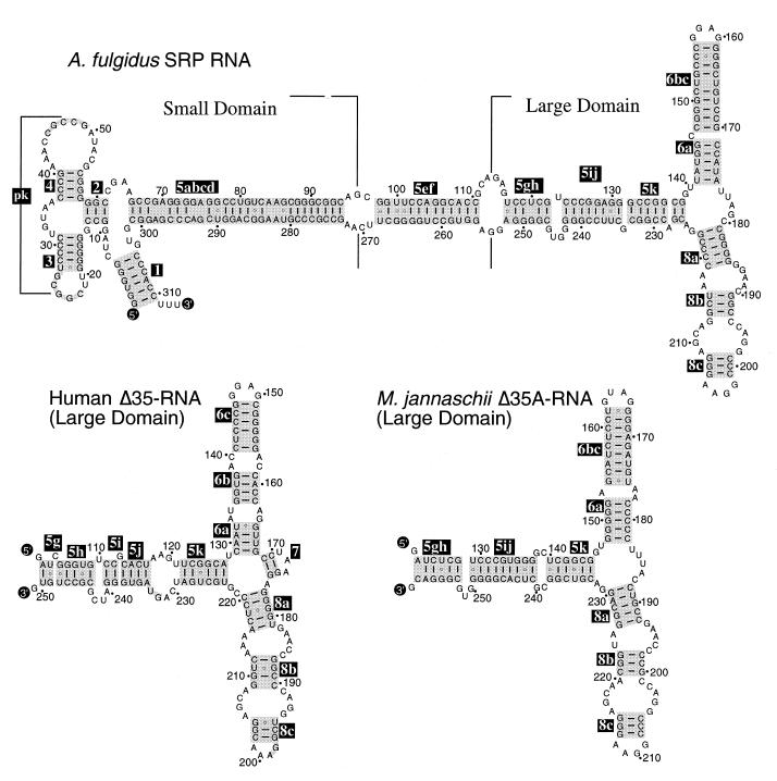
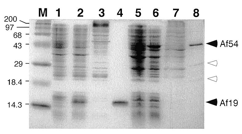
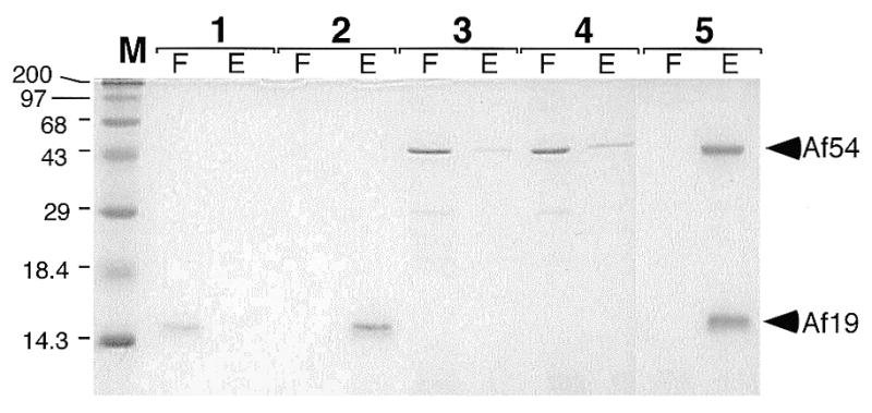
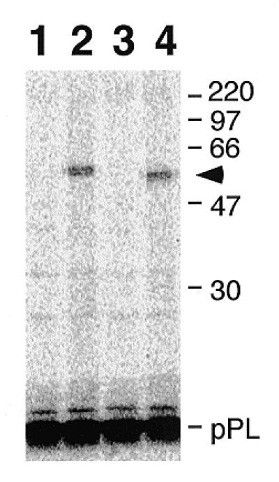
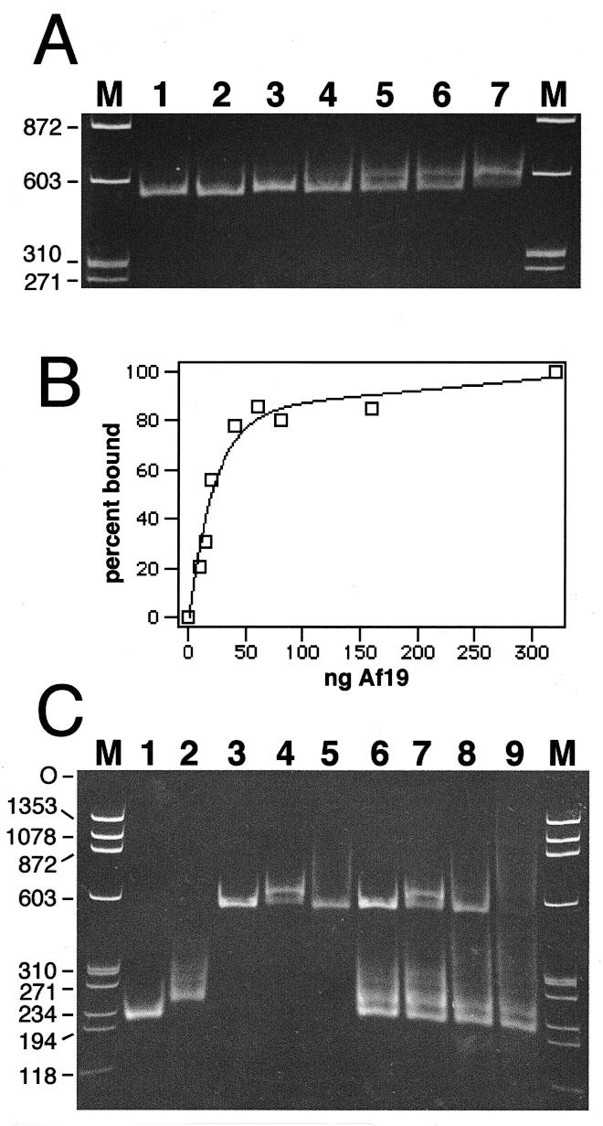
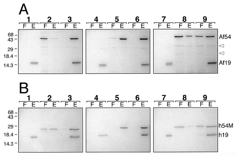
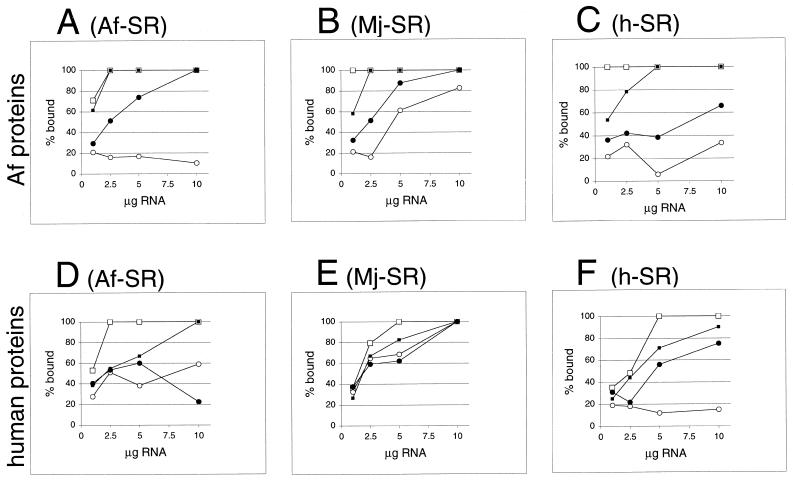
Signal recognition particle (SRP) takes part in protein targeting and secretion in all organisms. Searches for components of archaeal SRP in primary databases and completed genomes indicated that archaea possess only homologs of SRP RNA, and proteins SRP19 and SRP54. A recombinant SRP was assembled from cloned, expressed and purified components of the hyperthermophilic archaeon Archaeoglobus fulgidus. Recombinant Af-SRP54 associated with the signal peptide of bovine pre-prolactin translated in vitro. As in mammalian SRP, Af-SRP54 binding to Af-SRP RNA required protein Af-SRP19, although notable amounts bound in absence of Af-SRP19. Archaeoglobus fulgidus SRP proteins also bound to full-length SRP RNA of the archaeon Methanococcus jannaschii, to eukaryotic human SRP RNA, and to truncated versions which corresponded to the large domain of SRP. Dependence on SRP19 was most pronounced with components from the same species. Reconstitutions with heterologous components revealed a significant potential of human SRP proteins to bind to archaeal SRP RNAs. Surprisingly, M.jannaschii SRP RNA bound to human SRP54M quantitatively in the absence of SRP19. This is the first report of reconstitution of an archaeal SRP from recombinantly expressed purified components. The results highlight structural and functional conservation of SRP assembly between archaea and eucarya.


Figures

Figure 1
Figure 1
Secondary structures of A.fulgidus SRP RNA and Δ35 RNAs of H.sapiens and M.jannaschii. Secondary structures are shown with base pairings supported by comparative sequence analysis of SRP RNA sequences in the SRP database (14). 5′- and 3′-ends of RNA molecules are labeled as such; helices are numbered 2–8 according to the nomenclature of Larsen and Zwieb (25). Base paired sections of helices 5, 6 and 8, including regions of coaxial stacking (48), are highlighted in gray and labeled in reverse print with suffices a–k in helix 5, and a–c in helices 6 and 8. Residues are numbered in 10-nucleotide increments and marked with dots in 10-nucleotide increments in reference to full-length molecules.
Figure 2
Figure 2
Expression and purification of A.fulgidus SRP19 and SRP54. Proteins were separated on 15% SDS–polyacrylamide gels and stained with Coomassie blue. M, pre-stained high molecular weight markers (Gibco BRL) with sizes indicated in kDa. Lane 1, protein extract from uninduced E.coli BL21(DE3-pLysE) cells; lane 2, proteins from cells induced with IPTG for 2 h; lane 3, flowthrough from cation exchange chromatography; lane 4, pooled high-salt eluate; lane 5, protein extract from uninduced E.coli BL21(DE3-pSBETa) cells; lane 6, protein extract after induction with IPTG for 4 h; lane 7, flowthrough of Biorex 70 column; lane 8, pooled high-salt eluate. Solid arrowheads indicate positions of proteins Af-SRP54 (Af54) and Af-SRP19 (Af19), respectively. Open arrowheads mark Af-SRP54-derived polypeptides with predicted molecular weights of 30 359 and 17 855, likely a result of proteolytic cleavage likely occurring between the G- and M-domain of Af-SRP54 (29,43).
Figure 3
Figure 3
SRP RNA binding activities of Af-SRP19 and Af-SRP54 proteins. Proteins separated by electrophoresis on 15% polyacrylamide gels and stained with Coomassie blue in the flowthrough (F) and high-salt eluate (E) of DEAE-columns (see Materials and Methods). Lane 1, 1 µg Af-SRP19 in absence of Af-SRP RNA; lane 2, 1 µg Af-SRP19 mixed with 10 µg Af-SRP RNA; lane 3, 3.8 µg Af-SRP54 in absence of Af-SRP RNA; lane 4, 3.8 µg Af-SRP54 added to 10 µg Af-SRP RNA; lane 5, 1 µg Af-SRP19 and 3.8 µg Af-SRP54 with 10 µg of Af-SRP RNA. Pre-stained high molecular weight markers (Gibco BRL) with sizes indicated in kDa in lane M.
Figure 4
Figure 4
Photoaffinity labeling of purified Af-SRP54 with signal peptide. TDBA-modified lysine-tRNA was incorporated into the 86 amino acid residue bovine pre-prolactin (pPL) signal by translation in vitro (54) and used for photo cross-linking. The high-salt stripped nascent polypeptide–ribosome complex was mixed with either canine SRP54 or Af-SRP54 followed by centrifugation through a sucrose cushion. The radioactively labeled polypeptides were separated by SDS–PAGE on 12% gels and subjected to fluorography. Lane 1, non-irradiated pPL in the presence of canine SRP54; lane 2, irradiated pPL in the presence of canine SRP54; lane 3, non-irradiated pPL in the presence of recombinant Af-SRP54; lane 4, irradiated pPL in the presence of Af-SRP54. The arrowhead marks the position of Af-SRP54 cross-linked to the truncated (86 amino acid residues) pPL. Marker proteins with molecular weights in kDa are shown on the right.
Figure 5
Figure 5
Formation of A.fulgidus SRP by mobility shift. Electrophoresis of SRP RNA–protein complexes on 6% native polyacrylamide gels. The nucleic acids were stained with ethidium bromide. (A) Binding of purified Af-SRP19 to Af-SRP RNA. Each reaction contained 200 ng RNA. Protein was added at RNA/protein molar ratios of 0.082 (lane 2), 0.21 (lane 3), 0.41 (lane 4), 0.61 (lane 5), 0.82 (lane 6) and 1.6 (lane 7). The sizes of double-stranded DNA fragments (HaeIII-digested ΦX174 DNA) used for reference (lanes M) are indicated in base pairs on the left. (B) Quantitative analysis of Af-SRP19 binding to Af-SRP RNA by mobility shift as shown in (A). (C) Binding of Af-SRP19 and Af-SRP54 to various SRP RNAs. Lane 1, 200 ng Δ35-SRP RNA of M.jannaschii (Mj-Δ35); lane 2, 200 ng human Δ35-SRP RNA (h-Δ35); lane 3, 200 ng full-length A.fulgidus SRP RNA (Af-SRP); lane 4, 200 ng Af-SRP with 60 ng Af-SRP19 protein; lane 5, 200 ng Af-SRP with 700 ng Af-SRP54; lane 6, mixture of Mj-Δ35, h-Δ35 and Af-SRP RNA with amounts identical to those used in lanes 1–3; lane 7, RNA mixture as in lane 6 with the addition of 60 ng Af-SRP19; lane 8, RNA mixture as in lane 6 with the addition of 700 ng Af-SRP54; lane 9, RNA mixture as in lane 6 with addition of 60 ng Af-SRP19 and 700 ng Af-SRP54. Sizes of double-stranded DNA fragments used for reference (lanes M) are indicated in base pairs on the left.
Figure 6
Figure 6
Assembly with proteins of A.fulgidus or human SRP. (A) Binding of 1 µg Af-SRP19 and 3.8 µg Af-SRP54 polypeptides to 10 µg Af-SRP RNA (lanes 1–3), 10 µg Mj-SRP RNA (lanes 4–6) or 10 µg human SRP RNA (lanes 7–9). (B) Binding of 1.3 µg human SRP19 and 2 µg human SRP54M polypeptides to 10 µg Af-SRP RNA (lanes 1–3), 10 µg Mj-SRP RNA (lanes 4–6) or 10 µg human SRP RNA (lanes 7–9). Each panel shows polypeptides in the flowthrough (F) and eluate (E) as determined in DEAE binding assays (see Materials and Methods) followed by electrophoresis of polypeptides on 15% polyacrylamide gels and staining with Coomassie blue. Lanes 1, 4 and 7, addition of SRP19; lanes 2, 5 and 8, addition of Af-SRP54 or human SRP54M; lanes 3, 6 and 9, addition of both Af-SRP19 and Af-SRP54 (A) or human SRP19 and human SRP54M (B). Mobilities of Af-SRP54 (Af54), Af-SRP19 (Af19), human SRP19 (h19) and human SRP54M (h54M) polypeptides are indicated on the right. Open arrowheads in (A) mark two proteolytic products of Af-SRP54. Migration distances of molecular weight markers with sizes in kDa are indicated on the left.
Figure 7
Figure 7
Formation of ribonucleoprotein particles with A.fulgidus, M.jannaschii or human SRP RNAs. Activity of A.fulgidus SRP proteins (A–C) or human SRP proteins (D–F) with variable amounts of SRP RNAs of A.fulgidus (A) and (D), M.jannaschii (B) and (E) or H.sapiens (C) and (F) measured in the DEAE affinity assays (see Materials and Methods) from the intensity of Coomassie blue-stained polypeptides separated by SDS–PAGE as shown in Figure 6. Binding of SRP19 proteins is indicated by lines connected to open squares; open circles mark binding of Af-SRP54 (A–C) or human SRP54M (D–F). Solid squares indicate binding of SRP19 in presence of Af-SRP54 or human SRP54M; solid circles show binding of Af-SRP54 or human SRP54M in presence of SRP19. Small amounts of background binding observed without RNA or in presence of tRNA in the range of 3–10% observed with Af-SRP54 or human SRP54M (see Results) were plotted. Equimolar protein concentrations were 1 µg Af-SRP19, 3.8 µg Af-SRP54, 1.3 µg human SRP19 and 2 µg human SRP54M per 50 µl reaction volume.

References

    1. Lütcke H. (1995) Eur. J. Biochem., 228, 531–550. - PubMed
    1. Bui N. and Strub,K. (1999) Biol. Chem., 380, 135–145. - PubMed
    1. Walter P. and Blobel,G. (1981) J. Cell Biol., 91, 551–556. - PMC - PubMed
    1. Walter P., Ibrahimi,I. and Blobel,G. (1981) J. Cell Biol., 91, 545–550. - PMC - PubMed
    1. Walter P. and Blobel,G. (1981) J. Cell Biol., 91, 557–561. - PMC - PubMed

Publication types

MeSH terms