Comparative Study
. 2000 Mar 15;28(6):1397-406.
doi: 10.1093/nar/28.6.1397.

Genome sequences of Chlamydia trachomatis MoPn and Chlamydia pneumoniae AR39

Affiliations
Comparative Study

Genome sequences of Chlamydia trachomatis MoPn and Chlamydia pneumoniae AR39

T D Read et al. Nucleic Acids Res. .

Abstract

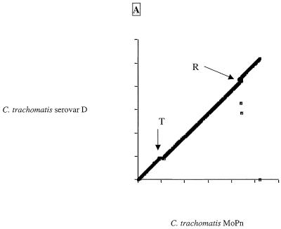
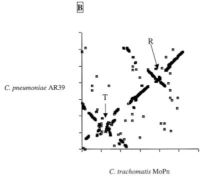
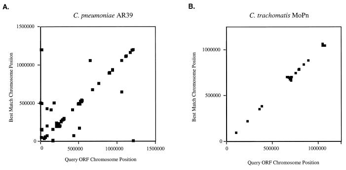
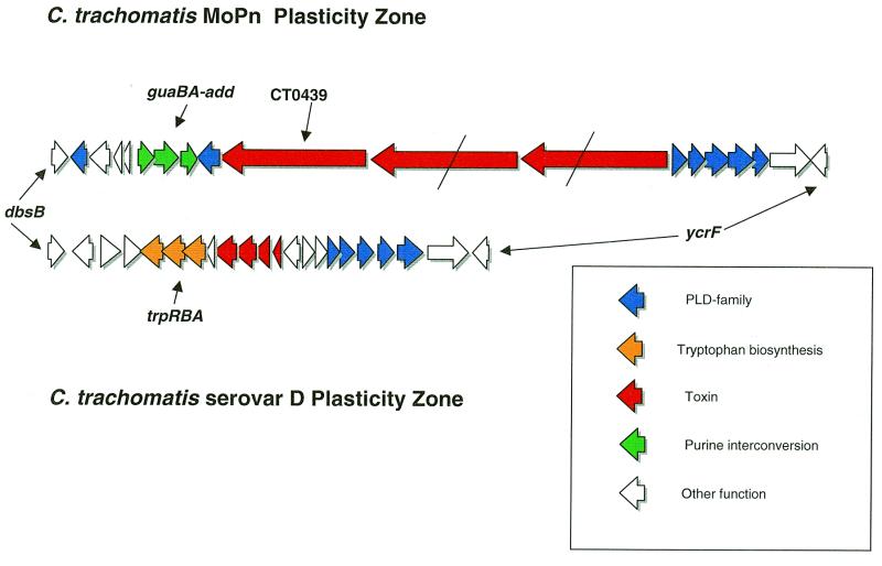
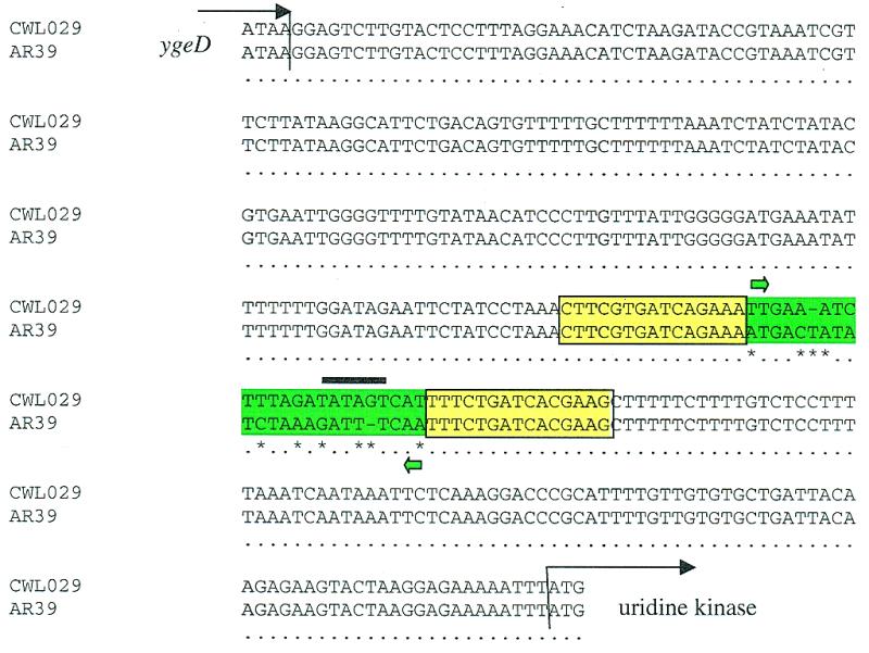
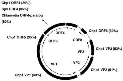
The genome sequences of Chlamydia trachomatis mouse pneumonitis (MoPn) strain Nigg (1 069 412 nt) and Chlamydia pneumoniae strain AR39 (1 229 853 nt) were determined using a random shotgun strategy. The MoPn genome exhibited a general conservation of gene order and content with the previously sequenced C.trachomatis serovar D. Differences between C.trachomatis strains were focused on an approximately 50 kb 'plasticity zone' near the termination origins. In this region MoPn contained three copies of a novel gene encoding a >3000 amino acid toxin homologous to a predicted toxin from Escherichia coli O157:H7 but had apparently lost the tryptophan biosyntheis genes found in serovar D in this region. The C. pneumoniae AR39 chromosome was >99.9% identical to the previously sequenced C.pneumoniae CWL029 genome, however, comparative analysis identified an invertible DNA segment upstream of the uridine kinase gene which was in different orientations in the two genomes. AR39 also contained a novel 4524 nt circular single-stranded (ss)DNA bacteriophage, the first time a virus has been reported infecting C. pneumoniae. Although the chlamydial genomes were highly conserved, there were intriguing differences in key nucleotide salvage pathways: C.pneumoniae has a uridine kinase gene for dUTP production, MoPn has a uracil phosphororibosyl transferase, while C.trachomatis serovar D contains neither gene. Chromosomal comparison revealed that there had been multiple large inversion events since the species divergence of C.trachomatis and C.pneumoniae, apparently oriented around the axis of the origin of replication and the termination region. The striking synteny of the Chlamydia genomes and prevalence of tandemly duplicated genes are evidence of minimal chromosome rearrangement and foreign gene uptake, presumably owing to the ecological isolation of the obligate intracellular parasites. In the absence of genetic analysis, comparative genomics will continue to provide insight into the virulence mechanisms of these important human pathogens.


Figures

Figure 1
Figure 1
Comparison of the C.pneumoniae and C.trachomatis genomes. The DNA genomes of C.trachomatis MoPn, C.trachomatis serovar D and C.pneumoniae (CWL029 and AR39 are effectively identical at this level of resolution) are represented as circles. Genomes are scaled in 100 000 nt increments. The three outer rings of each genome represent assignment of genes sharing identity in a Fasta3 comparison with a score of <0.00000001. Each tick indicates the location of the 5′-end of a gene. In the first, outer ring the green ticks represent genes encoding proteins conserved in all Chlamydia genomes, the purple ticks are genes conserved in C.pneumoniae and one C.trachomatis genome and are hence assumed to be deleted from the other C.trachomatis genome. The second circle (red) shows species-specific genes. The third circle (blue) illustrates genes encoding proteins not similar at a score of <10–8 to any other chlamydial protein. The fourth circle shows the location of the tRNAs and the fifth the position of the rRNA operons. The inner, sixth circle shows the results of GC skew analysis using a 1000 nt window size (12). Windows with a positive skew value are shown as cyan ticks, with a negative skew as yellow. The origin of replication and the termination region are defined as the points of inflection from positive to negative and negative to positive skew, respectively. The approximate positions of the plasticity zones near the origins of replication are indicated by the letters PZ. The genomes have been branched in a phylogenetic tree based on the average identity of homologous genes. Branch lengths (bracketed value) are the average percent difference in the homologous genes.
Figure 2
Figure 2
Dot plots of gene similarities between (A) C.trachomatis MoPn and C.trachomatis serovar D and (B) C.trachomatis MoPn and C.pneumoniae AR39. The criterion for match was a Fasta3 score of <10–8. The genomes have been rotated to better show inversion around the origins. Axes are marked with 200 kb gradations. R and T are the locations of the origin of replication and termination region, respectively.
Figure 2
Figure 2
Dot plots of gene similarities between (A) C.trachomatis MoPn and C.trachomatis serovar D and (B) C.trachomatis MoPn and C.pneumoniae AR39. The criterion for match was a Fasta3 score of <10–8. The genomes have been rotated to better show inversion around the origins. Axes are marked with 200 kb gradations. R and T are the locations of the origin of replication and termination region, respectively.
Figure 3
Figure 3
Gene duplications in (A) C.pneumoniae AR39 and (B) C.trachomatis MoPn. Duplicated genes in each chromosome were identified as having a score in a Fasta3 comparison of <10–8. The location of each gene and its duplicate on the chromosome were plotted.
Figure 4
Figure 4
Gene map of C.trachomatis MoPn and serovar D plasticity zones. Schematic diagram showing the gene content of the plasticity zones of the two C.trachomatis strains between the conserved dbsB and ycrfV loci. The line across the two homologs of the large toxin gene of MoPn TC0439 indicates a frameshift mutation. The ‘toxin’ genes of serovar D are homologous to regions of the larger TC0439 protein.
Figure 5
Figure 5
Regions upstream of the uridine kinase gene in C.pneumoniae CWL029 and AR39. A comparison of the sequence between ygeD and uridine kinase of the CWL029 and AR39 strains is shown. Differences between the sequences (asterisks) are limited to the 23 bp inverted sequence (green highlight). This inverted segment contains a putative –10 sequence (black bar) TATAGT in the same orientation as the uridine kinase gene in CWL029. Flanking the inverted sequence are 15 kb inverted repeats (boxed yellow).
Figure 6
Figure 6
Comparison of C.pneumoniae AR39 phage with other sequences. The figure details putative ORFs of the 4524 nt ssDNA phage sequence (inner circle) with similarities to previously sequenced proteins. Chp1, C.psittaci Chp1 bacteriophage; Spv, Spiroplasma virus 4.

References

    1. Moulder J.W. (1991) Microbiol. Rev., 55, 143–190. - PMC - PubMed
    1. McClarty G. (1994) Trends Microbiol., 2, 157–164. - PubMed
    1. Rasmussen S.J., Eckmann,L., Quayle,A.J., Shen,L., Zhang,Y.X., Anderson,D.J., Fierer,J., Stephens,R.S. and Kagnoff,M.F. (1997) J. Clin. Invest., 99, 77–87. - PMC - PubMed
    1. Stephens R.S., Kalman,S., Lammel,C., Fan,J., Marathe,R., Aravind,L., Mitchell,W., Olinger,L., Tatusov,R.L., Zhao,Q., Koonin,E.V. and Davis,R.W. (1998) Science, 282, 754–759. - PubMed
    1. Kalman S., Mitchell,W., Marathe,R., Lammel,C., Fan,J., Hyman,R.W., Olinger,L., Grimwood,J., Davis,R.W. and Stephens,R.S. (1999) Nature Genet., 21, 385–389. - PubMed

Publication types

MeSH terms

Associated data