

Review
doi: 10.1385/1-59259-684-3:535.
Use of electron microscopy in the detection of adrenergic receptors
Affiliations
- PMID: 10685434
- PMCID: PMC2882091
- DOI: 10.1385/1-59259-684-3:535
Item in Clipboard
Review
Use of electron microscopy in the detection of adrenergic receptors
Methods Mol Biol.
2000.
No abstract available
Figures
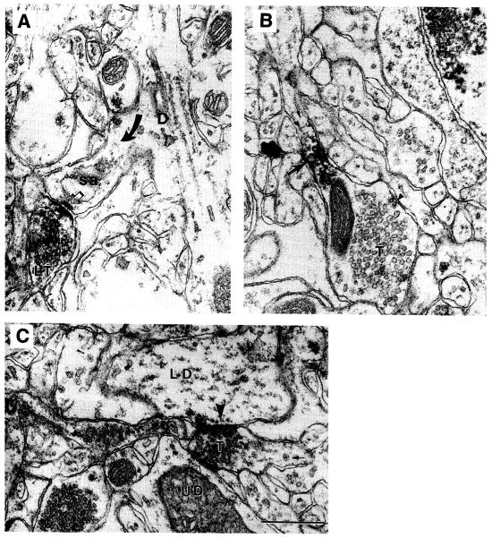
EMs obtained from the monkey prefrontal cortex immunolabeled using the α2A-AR antiserum and HRP-DAB as the immunolabel. (A) The receptor occurs directly over the presynaptic plasma membrane of a labeled terminal (small arrow in LT) forming a synaptic junction (open arrow) with a spine emanating from a dendritic shaft (arrow and D; sa = spine apparatus). (B) The receptor also occurs away from the synaptic region of a terminal, T, and instead at an intervaricose portion (large arrow and in between the two small arrows). The arrowhead points to a patch of plasma membrane undergoing endocytosis. (C) A terminal, T, forming a synaptic junction Simultaneously with two dendrites, one of which exhibits immunoreactivity over the postsynaptic membrane (LD, arrowhead) and another that is unlabeled (UD). The tissue was fixed by transcardial perfusion with a mixture of acrolein and paraformaldehyde, and then postfixed following the ICC procedure with osmium tetroxide. Calibration bar = 500 nm for all panels (from ref. , reproduced with permission from Oxford University Press).

EMs obtained from serially collected ultrathin sections of tissue immunolabeled dually for the catecholaminergic terminal (CT) marker, tyrosine hydroxylase, and the C-terminus of β-ARs are identified by the HRP-DAB label. β-ARs occur in fine astrocytic processes (β-A) that surround neurites, including one catecholaminergic terminal (CT) and three unlabeled terminals (uT1, uT2, and uT3). uT2 and uT3 are forming asymmetric synaptic junctions with dendritic spines (open arrows point to the thick postsynaptic densities), indicating that they utilize glutamate for excitatory synaptic transmission. An arrangement of this sort, whereby β-A occur inserted between CTs and excitatory synapses, is supportive of the idea that activated β-A enhance excitatory synaptic transmission by reducing astrocytic uptake of l -glutamate. Arrowhead pairs in panel A point to a gap junction formed between two β-As. Calibration bar = 500 nm. (From ref. , reproduced with permission from the Society for Neuroscience).

EMs obtained from postnatal d 10 rat visual cortex, showing β-AR immunoreactivity using HRP-DAB label and an antiserum directed against the C-terminus of the receptor. (A) A dendritic shaft receiving two synaptic inputs from unlabed axon terminals (T) one of which is immunolabeled over the postsynaptic density (curved arrow) and another that is unlabeled (open arrow points to the postsynaptic density).Arrowheads point to the unlabeled smooth saccules, indicating robust membrane turnover, that accompany the process of maturatlOn. Another profile,exhibits immunoreactivity along the plasma membrane: judging from its irregular contour (asterisks) it is most likely a glial process (LG). (B) The same antiserum β-ARs in presynaptic terminals (LT), identified by the clustering of small clear vesicles) (curved arrow) The same terminal contains a dense-cored vesicle (arrowhead). (A,C) β-AR immunoreactivity at postsynaptic sites occur not only over postsynaptic densities, but adjoining intracellular membranes and plasma membranes (small arrows). Calibration bar = 500 nm (from ref. , reproduced with permission from Cambridge University Press).

EMs revealing the differential localization of (β-ARs in astrocytes and neurons of adult visual cortex by SIG and their relation to GABA-immunoreactivity. (A) The dendrite from layer 5/6a of adult rat visual cortex exhibits numerous SIG particles (e.g., arrows), reflecting the presence of cytosolic (β-ARs. Within a neighboring astrocytic process (A), the SIG particles are close to the plasma membrane. Asterisks point to the irregular contours of the astrocyte. The same dendrite exhibits numerous lO-nm colloidal gold particles, resulting from EM-ICC detection of GABA, using PEG as label (e.g., Circled particles). A terminal, GT, contacting the dendrite exhibits high densitY of PEG labeling, indicating that it is a GABAergic terminal. (B) At a higher magnification, the neuropil from layer 2/3 of rat visual cortex exhibits five astrocytic processes (A1–A5). A1–A4 are immediately adjacent to asymmetric synaptic junctions (probably excitatory). A1, A3, and A5 exhibit robust (β-AR immunoreactivity (small arrows) primarily along the plasma membrane, but A2 and A4 exhibit much lower levels of (β-AR immunoreactivity and at sites away from the plasma membrane. Note that A4 is GABA-immunoreactive. In contrast, a dendritic process exhibits robust immunoreactivity for (β-ARs at sites away from the plasma membrane (e.g., arrow in (βD). A1 contacts an unlabeled terminal, UT, and also envelopes a dendrite, GD, identified to be GABAergic by the prevalence of PEG labels (circle), and synaptically associated with UT (curved arrow points to the postsynaptic density). Calibration bar = 500 nm.

An EM of the neuropil of an adult visual cortex, showing the coexistence of three antigens within a single dendrite. Triple EM-ICC was achieved by combining SIG to immunolabel NR2A subunits of NMDA-type glutamate receptors (circles), 30 nm colloidal gold-PEG for β-ARs (small arrows), and 10 nm colloidal gold-PEG for the inhibitoryneurotransmitter, GABA (arrowheads). This result indicates that a GABAergic inhibitory interneuron is receptive to noradrenaline as well as l -glutamate. The dendrite is also receptive to GABA, since it is forming a contact with two GABAergic terminals (T), one of which is associated with a morphologically identifiable synapse (open arrow points to the postsynaptic membrane). The localization of the two receptors away from the plasma membrane may be an indication that the receptors are in a desensitized state because of synaptic transmission that occurred during or prior to tissue fixation. Calibration bar = 500 nm.
References
-
- Strader CD, Fong TM, Graziano MP, Tota MR. The family of G-protein-coupled receptors. FASEB J. 1995;9:745–754. - PubMed
-
- Rohrer DK, Kobilka BK. Insights from in vivo modification of adrenergic receptor gene expression. Annu. Rev. Pharmacol. Toxicol. 1998;38:351–373. - PubMed
-
- Aoki C, Go CG, Venkatesan C, Kurose H. Perikaryal and synaptic localization of α2A-adrenergic receptor immunoreactivity in brain as revealed by light and electron microscopic immunocytochemistry. Brain Res. 1994;650:181–204. - PubMed
-
- Kalsner S, Westfall TC. Presynaptic receptors and the question of autoregulation of neurotransmitter release. Ann. NY Acad. Sci. 1990;604:652. - PubMed
-
- Venkatesan C, Kurose H, Aoki C. Cellular and subcellullar distribution of α2A- adrenergic receptor in the visual cortex of neonatal and adult rats. J. Comp. Neurol. 1996;365:79–95. - PubMed
Publication types
MeSH terms
Substances
Grants and funding
LinkOut - more resources
Full Text Sources
