. 2000 Mar;20(6):1982-92.
doi: 10.1128/MCB.20.6.1982-1992.2000.

An AU-rich sequence element (UUUN[A/U]U) downstream of the edited C in apolipoprotein B mRNA is a high-affinity binding site for Apobec-1: binding of Apobec-1 to this motif in the 3' untranslated region of c-myc increases mRNA stability

Affiliations

An AU-rich sequence element (UUUN[A/U]U) downstream of the edited C in apolipoprotein B mRNA is a high-affinity binding site for Apobec-1: binding of Apobec-1 to this motif in the 3' untranslated region of c-myc increases mRNA stability

S Anant et al. Mol Cell Biol. 2000 Mar.

Abstract

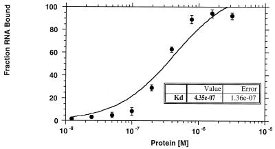
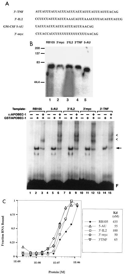
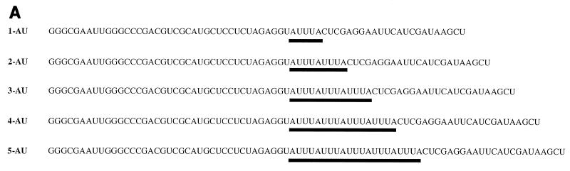
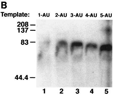
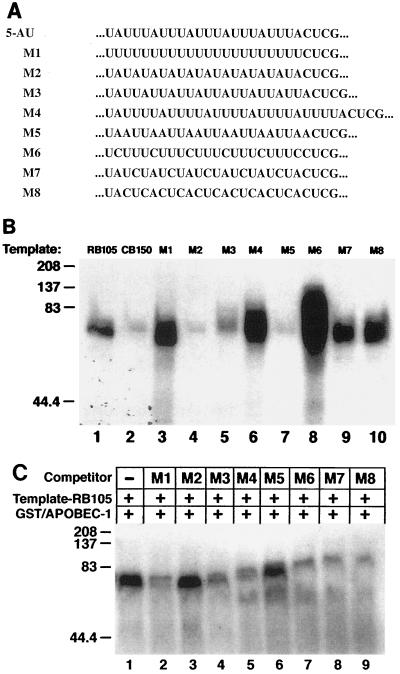
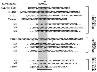
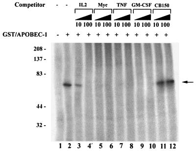
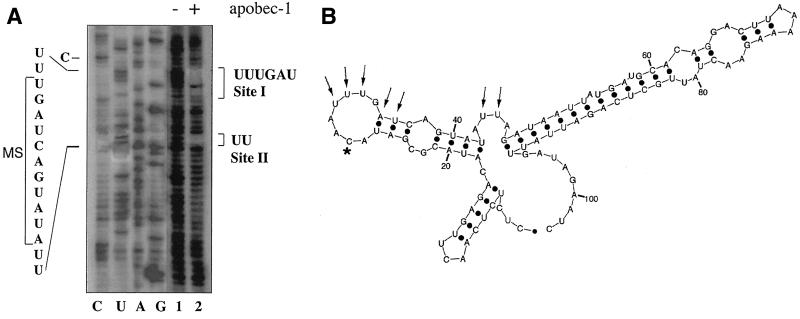
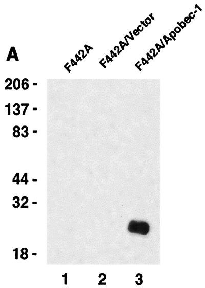
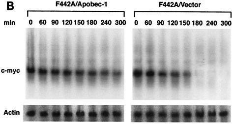
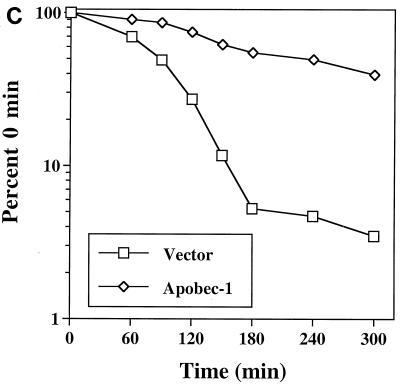
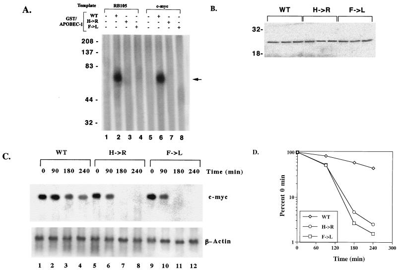
Apobec-1, the catalytic subunit of the mammalian apolipoprotein B (apoB) mRNA-editing enzyme, is a cytidine deaminase with RNA binding activity for AU-rich sequences. This RNA binding activity is required for Apobec-1 to mediate C-to-U RNA editing. Filter binding assays, using immobilized Apobec-1, demonstrate saturable binding to a 105-nt apoB RNA with a K(d) of approximately 435 nM. A series of AU-rich templates was used to identify a high-affinity ( approximately 50 nM) binding site of consensus sequence UUUN[A/U]U, with multiple copies of this sequence constituting the high-affinity binding site. In order to determine whether this consensus site could be functionally demonstrated from within an apoB RNA, circular-permutation analysis was performed, revealing one major (UUUGAU) and one minor (UU) site located 3 and 16 nucleotides, respectively, downstream of the edited base. Secondary-structure predictions reveal a stem-loop flanking the edited base with Apobec-1 binding to the consensus site(s) at an open loop. A similar consensus (AUUUA) is present in the 3' untranslated regions of several mRNAs, including that of c-myc, that are known to undergo rapid degradation. In this context, it is presumed that the consensus motif acts as a destabilizing element. As an independent test of the ability of Apobec-1 to bind to this sequence, F442A cells were transfected with Apobec-1 and the half-life of c-myc mRNA was determined following actinomycin D treatment. These studies demonstrated an increase in the half-life of c-myc mRNA from 90 to 240 min in control versus Apobec-1-expressing cells. Apobec-1 expression mutants, in which RNA binding activity is eliminated, failed to alter c-myc mRNA turnover. Taken together, the data establish a consensus binding site for Apobec-1 embedded in proximity to the edited base in apoB RNA. Binding to this site in other target RNAs raises the possibility that Apobec-1 may be involved in other aspects of RNA metabolism, independent of its role as an apoB RNA-specific cytidine deaminase.


Figures

FIG. 1
FIG. 1
Binding affinity of GST–Apobec-1 to rat apoB RNA. Increasing amounts of GST–Apobec-1 (1.2 × 10⁻⁸ to 3.2 × 10⁻⁶ M) were bound to 15,000 cpm of 32P-radiolabeled 105-nt rat apoB transcript (RB105) and incubated at room temperature for 20 min. The mixture was then filtered through a nitrocellulose membrane, and the retained material was analyzed by scintillation spectroscopy. The data are plotted as the fraction of RNA bound (mean ± standard deviation) to the indicated amount of GST–Apobec-1 (Protein [M]). Each point represents data from three independent experiments.
FIG. 2
FIG. 2
RNA-protein complex formation between GST–Apobec-1 and AU-rich sequences present in the 3′ UTRs of rapidly degraded RNAs. (A) AU-rich sequences present in 3′ UTRs of TNF-α, IL-2, GM-CSF, and c-myc. These sequences were cloned into plasmid pGem-3Zf(+), linearized, and transcribed in the presence of [α-32P]UTP with T7 RNA polymerase. (B) Top: UV cross-linking. Five hundred nanograms of GST–Apobec-1 was incubated with 50,000 cpm of the indicated radiolabeled transcript followed by incubation with RNase T1 and heparin, subjected to UV cross-linking for 1.5 min, and analyzed by SDS–10% PAGE. The migration of the molecular mass markers is shown. Bottom: EMSA. GST–Apobec-1 was incubated with the indicated 32P-labeled RNA templates in the presence (+) or absence (−) of affinity-purified rabbit anti (α)-Apobec-1 IgG, and the resulting complexes were analyzed by nondenaturing polyacrylamide gel electrophoresis. Migration of the free probe (F), the GST–Apobec-1–RB105 complex (arrow), and the supershifted bands (arrowheads) is indicated. (C) Affinity of GST–Apobec-1 for AU-rich templates. Increasing amounts of GST–Apobec-1 (1.2 × 10⁻⁸ to 3.2 × 10⁻⁶ M) were bound to 15,000 cpm of the indicated 32P-radiolabeled transcript followed by filtration through a nitrocellulose membrane, and the retained material was analyzed by scintillation spectroscopy. The data are plotted as the fraction of RNA bound (mean ± standard deviation) to the indicated amount of GST–Apobec-1 (protein [M]). Each point represents data from three independent experiments.
FIG. 3
FIG. 3
Two tandem repeats of AUUU sequence are required for Apobec-1 binding. (A) Radiolabeled cRNAs were prepared containing one to five copies of an AUUUA motif (labeled 1-AU to 5-AU) and used in the in vitro UV cross-linking assays with GST–Apobec-1. The AUUU repeat in each transcript is underlined. Except for variations in AUUU iterations, each transcript contained the same flanking nucleotide sequence. (B) GST–Apobec-1 was incubated with the radiolabeled transcripts, subjected to UV cross-linking, and analyzed by SDS–10% PAGE. The migration of molecular mass markers is indicated.
FIG. 3
FIG. 3
Two tandem repeats of AUUU sequence are required for Apobec-1 binding. (A) Radiolabeled cRNAs were prepared containing one to five copies of an AUUUA motif (labeled 1-AU to 5-AU) and used in the in vitro UV cross-linking assays with GST–Apobec-1. The AUUU repeat in each transcript is underlined. Except for variations in AUUU iterations, each transcript contained the same flanking nucleotide sequence. (B) GST–Apobec-1 was incubated with the radiolabeled transcripts, subjected to UV cross-linking, and analyzed by SDS–10% PAGE. The migration of molecular mass markers is indicated.
FIG. 4
FIG. 4
GST–Apobec-1 binding to different AU-rich templates as a means to identify a consensus Apobec-1 binding site. (A) The AUUU sequence in construct 5-AU (shown underlined in Fig. 3A) was replaced with the indicated cassette, labeled M1 to M8. The flanking sequences in the various transcripts were identical. (B) UV cross-linking experiments were performed with GST–Apobec-1 and the indicated radiolabeled M transcripts, and the complex was analyzed by SDS–10% PAGE. RB105 (105-nt rat apoB cRNA; positive control) and CB150 (150-nt chicken apoB RNA; negative control) templates were also used in the cross-linking reaction. The migration of molecular mass markers is shown on the left. (C) Competition by the indicated M transcripts for GST–Apobec-1 binding to rat apoB RNA. Excess unlabeled M template cRNA was added to binding reaction mixtures containing radiolabeled RB105 and GST–Apobec-1. Following incubation, the reaction mixture was subjected to UV cross-linking followed by separation by SDS–10% PAGE and autoradiography. The migration of the molecular mass markers is shown on the left. +, present; −, absent.
FIG. 5
FIG. 5
Identification of a consensus Apobec-1 binding motif. UV cross-linking experiments to determine GST–Apobec-1 binding activity were performed (as shown in Fig. 4), and the sequences were arranged based on cross-linking efficiency. A consensus binding motif (UUUN[A/U]U) was derived from the alignment (shaded area).
FIG. 6
FIG. 6
Multiple tandem repeats of the consensus sequence make up the Apobec-1 high-affinity binding site. UV cross-linking experiments were performed with GST–Apobec-1 and radiolabeled RB105 RNA in the presence of (+) of 10- or 100-fold excess of unlabeled c-myc (Myc), TNF-α (TNF), IL-2 (IL-2), GM-CSF, or chicken apoB (CB150) transcripts and subjected to UV cross-linking. Following cross-linking, the reaction mixture was separated in an SDS–10% PAGE and autoradiographed. The migration of molecular mass markers (kilodaltons) is shown on the left. The location of the GST–Apobec-1–RNA cross-linked complex is indicated by an arrow. This is a representation of two independent tests.
FIG. 7
FIG. 7
Direct determination of Apobec-1 binding sites in rat apoB RNA. (A) A circular-permutation assay (CPA) was performed with GST–Apobec-1 and a 105-nt RB105 cRNA. RB105 RNA was circularized and subjected to partial alkaline hydrolysis under denaturing conditions to generate a complete population of linear circular-permutated (CP) isoforms. The CP isomers were mixed with GST–Apobec-1, and the bound isomers were recovered following filtration through a nitrocellulose membrane. Bound RNAs were identified by primer extension with Moloney murine leukemia virus reverse transcriptase using a 32P-labeled primer located at the 3′ end of the apoB sequence. Total CP isomers (lane 1) and GST–Apobec-1-bound isomers (lane 2), along with reverse transcriptase sequencing of the circular form of RB105 (lanes C, U, A, and G), were separated in an 8 M urea–8% polyacrylamide gel and autoradiographed. The sequence of interest is shown on the left, and the 11-nt mooring sequence motif (MS) is bracketed. The two Apobec-1 binding sites (sites I and II) are shown on the right. (B) Schematic representation of structure of a 105-nt apoB RNA, including the binding sites for Apobec-1 (determined by CPA). RNA folding was determined by using the RNA mfold program. RB105 forms a three-branch structure, and the Watson-Crick (A:U and G:C) and the weaker G:U wobble pairs are shown as black dots. Nucleotides required for Apobec-1 binding and the edited C are shown by arrows and an asterisk, respectively.
FIG. 8
FIG. 8
Binding of Apobec-1 to AU-rich sequence in 3′ UTR of c-myc mRNA increases c-myc mRNA stability. (A) Wild-type Apobec-1 cDNA was transfected into F442A cells, and stable transfectants were selected with G418 (F442A/apobec-1). As a control, vector alone was transfected and colonies were isolated (F442A/vector). Cytosolic S100 extracts were subjected to 30% ammonium sulfate precipitation, and aliquots were separated in an SDS–12% PAGE and blotted on a polyvinylidine difluoride membrane. The blot was probed with affinity-purified rabbit anti-Apobec-1 IgG, and bands were visualized by enhanced chemiluminescence. Molecular mass markers (kilodaltons) are indicated on the left. (B) Northern blot analysis of c-myc mRNA. Cells were grown to ∼90% confluence, and actinomycin D (final concentration, 10 μg/ml) was added. At the indicated time points, RNA was extracted, size separated in a formaldehyde-agarose gel, and blotted onto Nytran-Plus membranes. The blots were probed sequentially with c-myc and mouse β-actin cDNAs. (C) Hybridization was quantitated with a PhosphorImager and normalized to β-actin. Data from five independent experiments were averaged (mean ± standard deviation) and are presented as a percentage of c-myc mRNA remaining, relative to that at time zero. Error bars fall within the symbol for the mean.
FIG. 8
FIG. 8
Binding of Apobec-1 to AU-rich sequence in 3′ UTR of c-myc mRNA increases c-myc mRNA stability. (A) Wild-type Apobec-1 cDNA was transfected into F442A cells, and stable transfectants were selected with G418 (F442A/apobec-1). As a control, vector alone was transfected and colonies were isolated (F442A/vector). Cytosolic S100 extracts were subjected to 30% ammonium sulfate precipitation, and aliquots were separated in an SDS–12% PAGE and blotted on a polyvinylidine difluoride membrane. The blot was probed with affinity-purified rabbit anti-Apobec-1 IgG, and bands were visualized by enhanced chemiluminescence. Molecular mass markers (kilodaltons) are indicated on the left. (B) Northern blot analysis of c-myc mRNA. Cells were grown to ∼90% confluence, and actinomycin D (final concentration, 10 μg/ml) was added. At the indicated time points, RNA was extracted, size separated in a formaldehyde-agarose gel, and blotted onto Nytran-Plus membranes. The blots were probed sequentially with c-myc and mouse β-actin cDNAs. (C) Hybridization was quantitated with a PhosphorImager and normalized to β-actin. Data from five independent experiments were averaged (mean ± standard deviation) and are presented as a percentage of c-myc mRNA remaining, relative to that at time zero. Error bars fall within the symbol for the mean.
FIG. 9
FIG. 9
RNA binding mutants of Apobec-1 do not stabilize c-myc mRNA. (A) Wild-type (WT) and mutant (H→R and F→L) Apobec-1 cDNAs were expressed as GST fusion proteins and affinity purified over glutathione-agarose beads. Two hundred fifty nanograms of the indicated fusion protein was incubated with either radiolabeled RB105 or c-myc transcript, subjected to UV cross-linking, and analyzed by SDS–10% PAGE. The location of the GST–Apobec-1–RNA cross-linking complex is indicated by an arrow. The migration of molecular mass markers (kilodaltons) is indicated. This is a representation of two independent tests. (B) WT and mutant (H→R and F→L) Apobec-1 cDNAs were transiently transfected into F442A cells. Total cellular extracts were prepared, and aliquots were separated in an SDS–12% PAGE and transferred to a polyvinylidine difluoride membrane. The blot was probed with affinity-purified rabbit anti-Apobec-1 IgG, and the bands were visualized by enhanced chemiluminescence. The migration of molecular mass markers (kilodaltons) is indicated on the left. (C) WT and mutant (H→R and F→L) Apobec-1 cDNAs were transfected into F442A cells. Seventy-two hours after transfection, actinomycin D was added, the cells were incubated for the indicated times, and RNA was isolated. The RNA was subjected to Northern blot hybridization and probed with mouse c-myc and β-actin cDNAs. (D) The blots were scanned by PhosphorImager, and hybridization to the c-myc transcript was normalized to that of β-actin. The data from three independent transfections were averaged (mean ± standard deviation) and are presented as a percentage of c-myc mRNA remaining, relative to that at time zero. Error bars fall within the symbol for the mean.

References

    1. Anant S, MacGinnitie A J, Davidson N O. apobec-1, the catalytic subunit of the mammalian apolipoprotein B mRNA editing enzyme, is a novel RNA-binding protein. J Biol Chem. 1995;270:14762–14767. - PubMed
    1. Anant S, MacGinnitie A J, Davidson N O. The binding of apobec-1 to mammalian apo B RNA is stabilized by the presence of complementation factors which are required for posttranscriptional editing. Nucleic Acids Symp Ser. 1995;33:99–102. - PubMed
    1. Backus J W, Schock D, Smith H C. Only cytidines 5′ of the apolipoprotein B mRNA mooring sequence are edited. Biochim Biophys Acta. 1994;1219:1–14. - PubMed
    1. Backus J W, Smith H C. Three distinct RNA sequence elements are required for efficient apolipoprotein B (apoB) RNA editing in vitro. Nucleic Acids Res. 1992;20:6007–6014. - PMC - PubMed
    1. Bhattacharya S, Navaratnam N, Morrison J R, Scott J, Taylor W R. Cytosine nucleoside/nucleotide deaminases and apolipoprotein B mRNA editing. Trends Biochem Sci. 1994;19:105–106. - PubMed

Publication types