. 2000 Mar;20(6):2055-65.
doi: 10.1128/MCB.20.6.2055-2065.2000.

Disassociation of met-mediated biological responses in vivo: the natural hepatocyte growth factor/scatter factor splice variant NK2 antagonizes growth but facilitates metastasis

Affiliations

Disassociation of met-mediated biological responses in vivo: the natural hepatocyte growth factor/scatter factor splice variant NK2 antagonizes growth but facilitates metastasis

T Otsuka et al. Mol Cell Biol. 2000 Mar.

Abstract

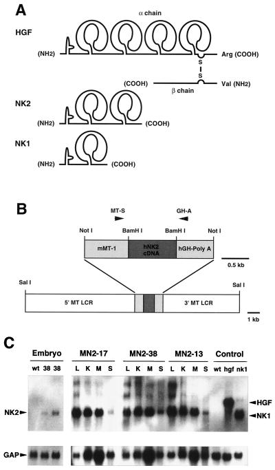
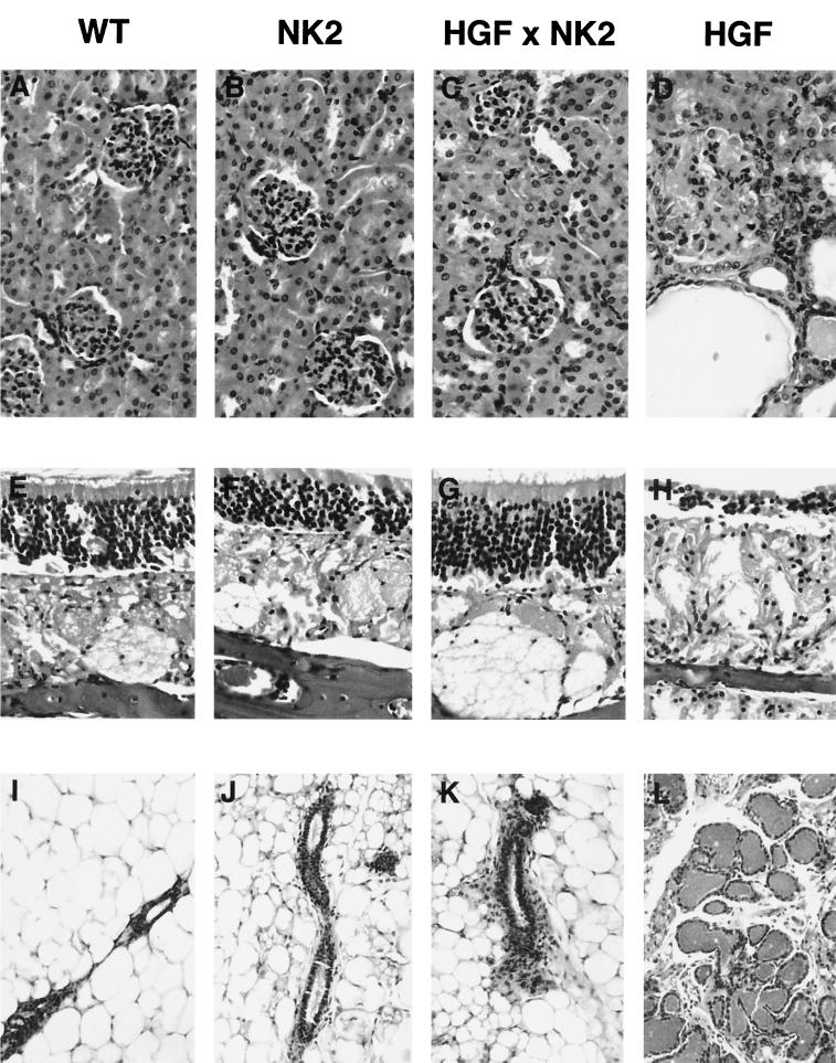
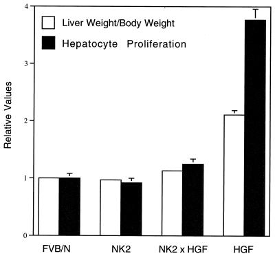
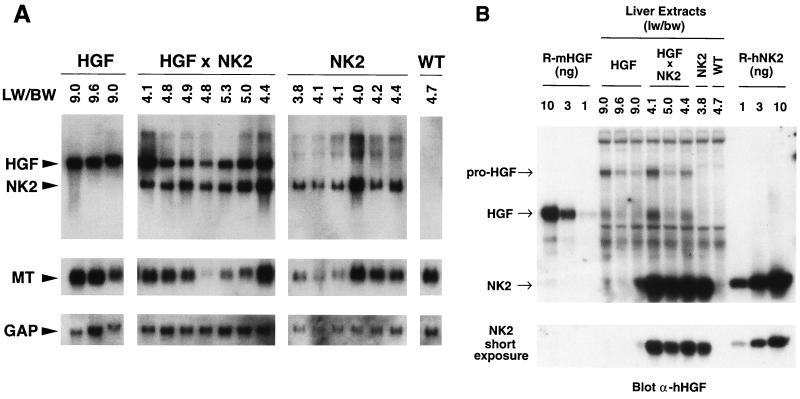
Hepatocyte growth factor/scatter factor (HGF/SF) stimulates numerous cellular activities capable of contributing to the metastatic phenotype, including growth, motility, invasiveness, and morphogenetic transformation. When inappropriately expressed in vivo, an HGF/SF transgene induces numerous hyperplastic and neoplastic lesions. NK1 and NK2 are natural splice variants of HGF/SF; all interact with a common receptor, Met. Although both agonistic and antagonistic properties have been ascribed to each isoform in vitro, NK1 retains the full spectrum of HGF/SF-like activities when expressed as a transgene in vivo. Here we report that transgenic mice broadly expressing NK2 exhibit none of the phenotypes characteristic of HGF/SF or NK1 transgenic mice. Instead, when coexpressed in NK2-HGF/SF bitransgenic mice, NK2 antagonizes the pathological consequences of HGF/SF and discourages the subcutaneous growth of transplanted Met-containing melanoma cells. Remarkably, the metastatic efficiency of these same melanoma cells is dramatically enhanced in NK2 transgenic host mice relative to wild-type recipients, rivaling levels achieved in HGF/SF and NK1 transgenic hosts. Considered in conjunction with reports that in vitro NK2 induces scatter, but not other activities, these data strongly suggest that cellular motility is a critical determinant of metastasis. Moreover, our results demonstrate how alternatively structured ligands can be exploited in vivo to functionally dissociate Met-mediated activities and their downstream pathways.


Figures

FIG. 1
FIG. 1
Structure and expression of NK2. (A) Schematic comparison of HGF/SF (designated as HGF in this and all other figures) and its natural splice variants NK2 and NK1. Each isoform contains a single so-called N domain at the amino terminus, and either four, two, or one kringle domain, as shown. However, only HGF/SF is processed into two chains, the β chain containing an enzymatically inactive serine protease domain. (B) The SalI-SalI NK2 transgene construct contained the human NK2 cDNA, the mouse MT gene promoter (mMT-1) and 5′ and 3′ flanking sequences (MT LCR), and the hGH poly(A) signal. Mice harboring the NK2 transgene were identified by PCR using primers MT-S and GH-A, as indicated. (C) Analysis of NK2 transgene expression in mouse tissues by Northern blot hybridization. For embryonic expression (three-lane panel at left), wild-type (wt) embryos were harvested at E16.5, and transgenic embryos from line MN2-38 (38) were harvested at E14.5 (middle lane) and E16.5 (right lane). Adult (2-month-old) tissues from three independently generated lines, MN2-17, MN2-38, and MN2-13, were studied. Tissues analyzed included liver (L), kidney (K), skeletal muscle (M), and skin (S). The control lanes at far right show expression of HGF/SF sequences in livers of wild-type and HGF/SF and NK1 transgenic mice. Following hybridization with a human NK2 cDNA probe (top panels), the filter was stripped and rehybridized with a control GAP cDNA probe (bottom panels).
FIG. 2
FIG. 2
NK2 extinguishes the phenotypic consequences of ectopic HGF/SF expression in bitransgenic mice. Shown are panels of tissues, including kidney (A to D), olfactory mucosa (E to H), and virgin mammary gland (I to L), from wild-type (A, E, and I), NK2 transgenic (B, F, and J), HGF/SF-NK2 bitransgenic (C, G, and K), and HGF/SF transgenic (D, H, and L) mice. Tissues shown are from mice 2.5 months of age. Note that bitransgenic tissues resemble wild-type tissues and do not contain the pathological features characteristically evident in HGF/SF transgenic animals.
FIG. 3
FIG. 3
NK2 inhibits the proliferative effects of HGF/SF on hepatocytes. Shown are mean liver weight/body weight ratios (white bars) and hepatocyte labeling indices (black bars) from wild-type (FVB/N), NK2 transgenic, NK2-HGF/SF bitransgenic, and HGF/SF transgenic livers. All values were normalized to wild type (set at 1.0), and error bars represent standard errors of the means. For both liver size and hepatocyte proliferation, the P value is <0.0001 in NK2-HGF/SF bitransgenic versus HGF/SF transgenic mice.
FIG. 4
FIG. 4
Comparative transgene expression in livers of HGF/SF and NK2 transgenic and HGF/SF-NK2 bitransgenic mice. (A) Northern blot analysis of total liver RNA (15 μg/sample) using as probe mouse sequences equivalent to NK2 (top panels), mouse MT cDNA (middle panels), or mouse GAP cDNA (bottom panels). Numbers at the top indicate liver weight/body weight ratios (LW/BW). All mice were 1.5 months of age. (B) Quantitative Western blot analysis of mouse HGF/SF and human NK2 transgenic mouse liver extracts (25 μg/sample) using an anti-human HGF/SF antibody. Arrows mark positions of unprocessed pro-HGF/SF, processed HGF/SF, and NK2. Liver weight/body weight ratios (lw/bw) are shown at top. Recombinant mouse HGF/SF (R-mHGF) and human NK2 (R-hNK2) standards of known amounts (in nanograms) are displayed on the extreme left and right, respectively. A short exposure shown at bottom permits quantification of NK2 levels in liver extracts. In general, RNA and protein results in panels A and B, respectively, are in accord.
FIG. 5
FIG. 5
Quantification of Met and Met activity in melanoma cells. Extracts prepared from 37-32 melanoma cells treated with either nothing (control), HGF/SF, or NK2 were immunoprecipitated (IP) with anti-Met antibody and subsequently probed with either anti-phosphotyrosine (α-pY, top left) or anti-Met (α-c-Met, bottom left) antibodies. Extracts were also directly probed with anti-phospho-MAPK antibody (right). Molecular masses in kilodaltons are shown. Note that, relative to NK2, HGF/SF induces phosphorylation of Met and MAPK without altering the levels of Met.
FIG. 6
FIG. 6
NK2 antagonizes paracrine, but not autocrine, HGF/SF-induced subcutaneous melanoma growth. One million 37-32 melanoma cells were injected under the back skin of 2- to 3-month-old wild-type (WT), HGF/SF transgenic, NK2 transgenic, and HGF/SF-NK2 bitransgenic mice; tumor sizes were measured; and growth rates were calculated.
FIG. 7
FIG. 7
NK2 enhances metastatic efficiency, but not growth, of high-Met-expressing melanoma cells. The figure shows results of analysis of liver metastasis of melanoma cells in genetically modified host mice. One million 37-32 melanoma cells were injected intravenously into the tail vein of wild-type (WT), NK2 transgenic, NK1 transgenic, and HGF/SF transgenic mice. (A and B) After 3 weeks, livers were examined grossly (A) and histopathologically (B) for the presence of metastatic tumors. Melanomas were immunohistochemically visualized (brown staining) using an anti-mouse TRP1 antibody. (C) Liver preparations from the genetically modified host mice were used to quantify both mean numbers of 37-32 melanoma cell metastases (white bars) and mean tumor sizes (black bars). Error bars indicate standard errors of the means. There was no statistically significant difference in the numbers of metastases per liver in the three transgenic lines. For mean tumor size, P value was <0.001 for NK2 versus either NK1 or HGF/SF; P value was 0.2 for NK1 versus HGF/SF.
FIG. 7
FIG. 7
NK2 enhances metastatic efficiency, but not growth, of high-Met-expressing melanoma cells. The figure shows results of analysis of liver metastasis of melanoma cells in genetically modified host mice. One million 37-32 melanoma cells were injected intravenously into the tail vein of wild-type (WT), NK2 transgenic, NK1 transgenic, and HGF/SF transgenic mice. (A and B) After 3 weeks, livers were examined grossly (A) and histopathologically (B) for the presence of metastatic tumors. Melanomas were immunohistochemically visualized (brown staining) using an anti-mouse TRP1 antibody. (C) Liver preparations from the genetically modified host mice were used to quantify both mean numbers of 37-32 melanoma cell metastases (white bars) and mean tumor sizes (black bars). Error bars indicate standard errors of the means. There was no statistically significant difference in the numbers of metastases per liver in the three transgenic lines. For mean tumor size, P value was <0.001 for NK2 versus either NK1 or HGF/SF; P value was 0.2 for NK1 versus HGF/SF.

Similar articles

Cited by

References

    1. Bardelli A, Basile M L, Audero E, Giordano S, Wennstrom S, Menard S, Comoglio P M, Ponzetto C. Concomitant activation of pathways downstream of Grb2 and PI 3-kinase is required for MET-mediated metastasis. Oncogene. 1999;18:1139–1146. - PubMed
    1. Bellusci S, Moens G, Gaudino G, Comoglio P, Nakamura T, Thiery J-P, Jouanneau J. Creation of a hepatocyte growth factor/scatter factor autocrine loop in carcinoma cells induces invasive properties associated with increased tumorigenicity. Oncogene. 1994;9:1091–1099. - PubMed
    1. Bladt F, Riethmacher D, Isenmann S, Aguzzi A, Birchmeier C. Essential role for the c-met receptor in the migration of myogenic precursor cells into the limb bud. Nature. 1995;376:768–771. - PubMed
    1. Bottaro D P, Rubin J S, Faletto D L, Chan A M, Kmiecik T E, Vande Woude G F, Aaronson S A. Identification of the hepatocyte growth factor receptor as the c-met proto-oncogene product. Science. 1991;251:802–804. - PubMed
    1. Castagnino P, Soriano J V, Montesano R, Bottaro D P. Induction of tissue inhibitor of metalloproteinases-3 is a delayed early cellular response to hepatocyte growth factor. Oncogene. 1998;17:481–492. - PubMed

LinkOut - more resources