. 2000 Mar;20(6):2108-21.
doi: 10.1128/MCB.20.6.2108-2121.2000.

Cloning of a mammalian transcriptional activator that binds unmethylated CpG motifs and shares a CXXC domain with DNA methyltransferase, human trithorax, and methyl-CpG binding domain protein 1

Affiliations

Cloning of a mammalian transcriptional activator that binds unmethylated CpG motifs and shares a CXXC domain with DNA methyltransferase, human trithorax, and methyl-CpG binding domain protein 1

K S Voo et al. Mol Cell Biol. 2000 Mar.

Abstract

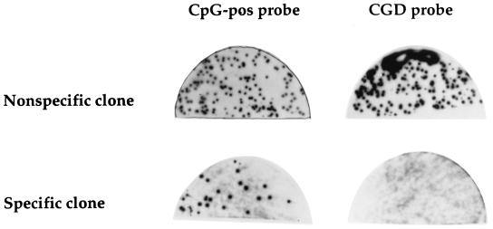
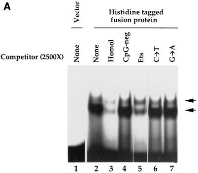
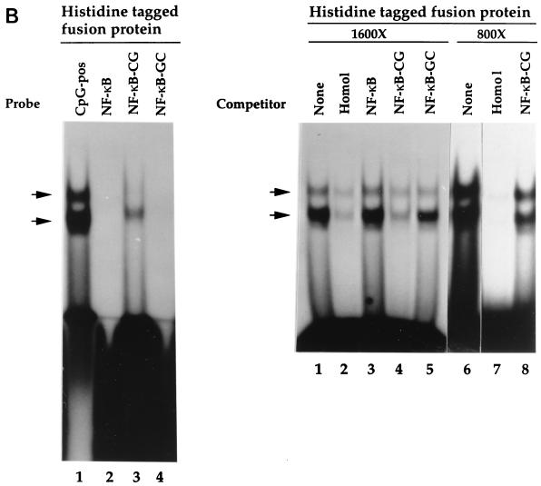
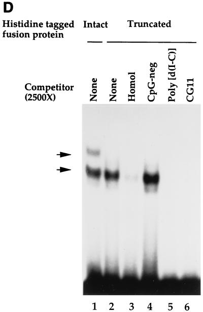
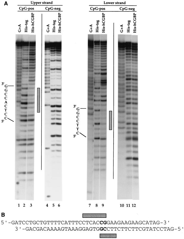
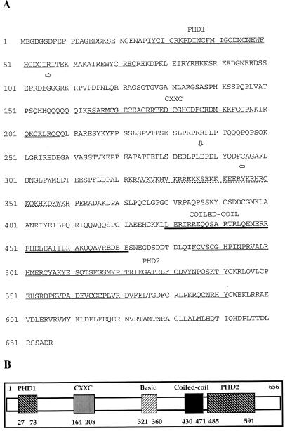
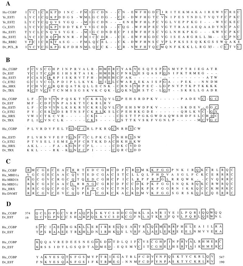
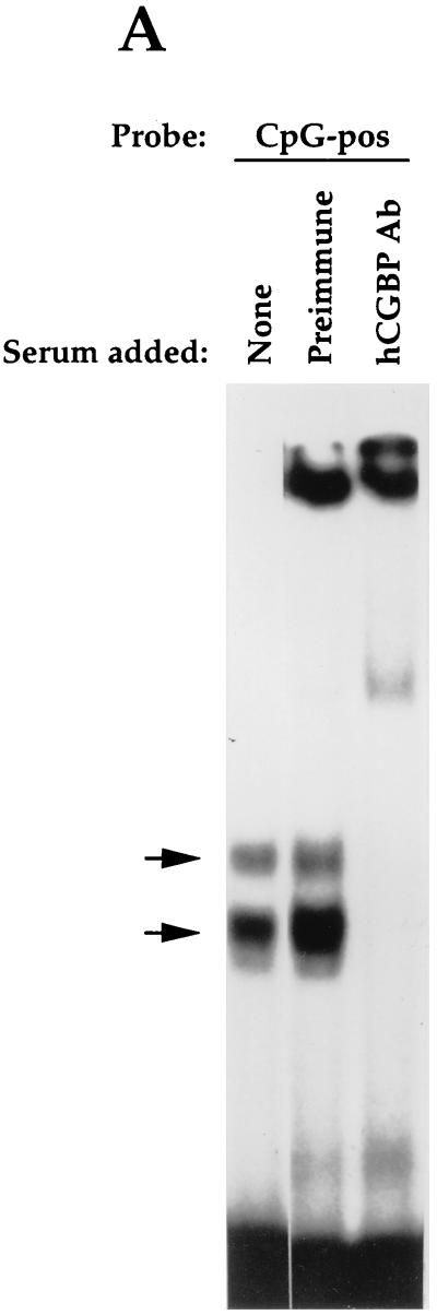
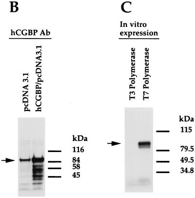
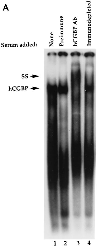
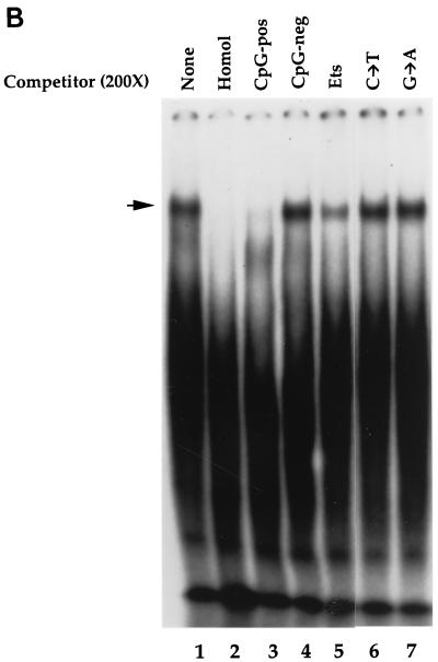
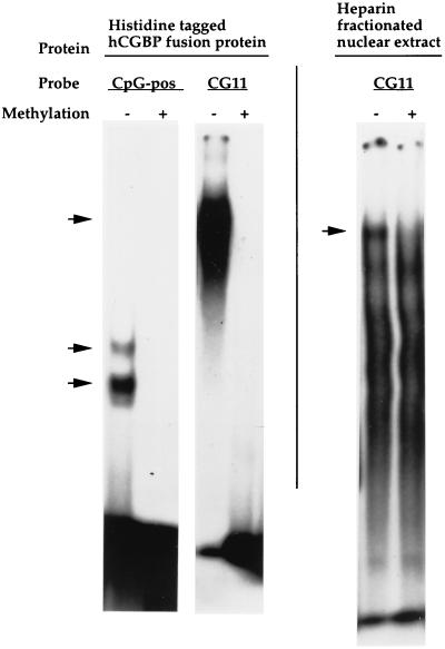
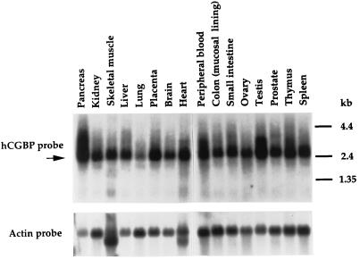
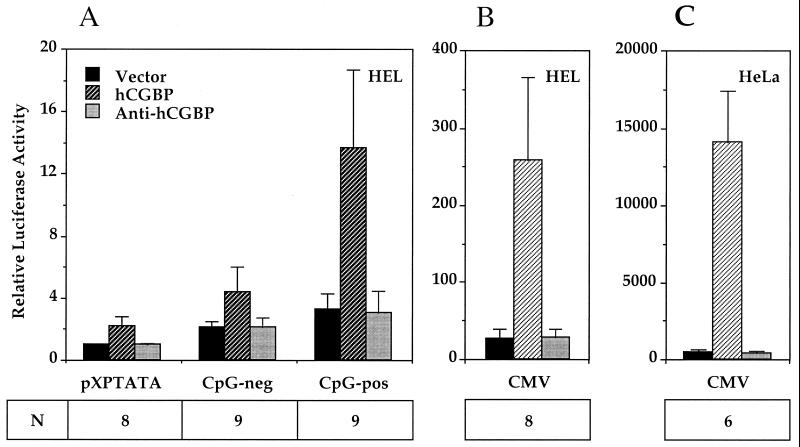
Ligand screening was utilized to isolate a human cDNA that encodes a novel CpG binding protein, human CpG binding protein (hCGBP). This factor contains three cysteine-rich domains, two of which exhibit homology to the plant homeodomain finger domain. A third cysteine-rich domain conforms to the CXXC motif identified in DNA methyltransferase, human trithorax, and methyl-CpG binding domain protein 1. A fragment of hCGBP that contains the CXXC domain binds to an oligonucleotide probe containing a single CpG site, and this complex is disrupted by distinct oligonucleotide competitors that also contain a CpG motif(s). However, hCGBP fails to bind oligonucleotides in which the CpG motif is either mutated or methylated, and it does not bind to single-stranded DNA or RNA probes. Furthermore, the introduction of a CpG dinucleotide into an unrelated oligonucleotide sequence is sufficient to produce a binding site for hCGBP. Native hCGBP is detected as an 88-kDa protein by Western analysis and is ubiquitously expressed. The DNA-binding activity of native hCGBP is apparent in electrophoretic mobility shift assays, and hCGBP trans-activates promoters that contain CpG motifs but not promoters in which the CpG is ablated. These data indicate that hCGBP is a transcriptional activator that recognizes unmethylated CpG dinucleotides, suggesting a role in modulating the expression of genes located within CpG islands.


Figures

FIG. 1
FIG. 1
Isolation by ligand screening of cDNA clones that encode DNA-binding factors. Phages derived from an HL60 λgt11 expression library were induced to express fusion proteins and incubated with CpG-pos (20) or CGD oligonucleotide probe (40) as described in Materials and Methods. Shown are a representative phage clone that encodes a non-sequence-specific DNA-binding activity (top) and a representative phage clone that exhibits sequence-specific DNA-binding activity (bottom).
FIG. 2
FIG. 2
The histidine-tagged fusion protein exhibits sequence-specific DNA-binding activity. (A) Binding specificity of the histidine-tagged fusion protein. EMSA was performed as described in Materials and Methods, using purified histidine-tagged fusion protein (720-bp cDNA fragment) and the CpG-pos probe. Competitor oligonucleotides were added as indicated. Lane 1, vector alone; lanes 2 to 7, addition of histidine-tagged fusion protein (lane 2, no competitor; lane 3, homologous competitor; lane 4, CpG-neg competitor [40]; lane 5, Ets oligonucleotide competitor (which contains a CpG motif); lane 6, mutated Ets oligonucleotide competitor in which the cytosine of the CpG motif is mutated to a thymine [C→T]; lane 7, same as lane 6, except the guanine of the CpG motif is mutated to adenine [G→A]). (B) Introduction of a CpG motif into an unrelated oligonucleotide is sufficient to produce a binding site for the histidine-tagged fusion protein. EMSA was performed as described for panel A. (Left panel) Lane 1, CpG-pos probe; lane 2, NF-κB probe; lane 3, NF-κB–CG probe; lane 4, NF-κB–GC probe. (Right panel) Competitor oligonucleotides (1,600- or 800-fold molar excess) were added to samples containing the CpG-pos probe. Lanes 1 and 6, no competitor; lanes 2 and 7, homologous competitor; lane 3, NF-κB competitor; lanes 4 and 8, NF-κB–CG competitor; lane 5, NF-κB–GC competitor. (C) The histidine-tagged fusion protein fails to interact with single-stranded nucleic acids. EMSA was performed as described for panel A. (Left panel) Lane 1, double-stranded CpG-pos probe; lane 2, single-stranded CpG-pos probe (upper strand); lane 3, single-stranded CpG-pos probe (lower strand); lane 4, RNA CpG-pos probe (upper strand). (Right panel) EMSA was performed with the CpG-pos probe and the following oligonucleotide competitors: lane 1, no competitor; lane 2, homologous competitor; lane 3, single-stranded CpG-pos (upper strand); lane 4, single-stranded CpG-pos (lower strand); and lane 5, RNA CpG-pos (upper strand). (D) A truncated histidine-tagged fusion protein retains DNA-binding activity. EMSA was performed with the CpG-pos probe, the truncated histidine-tagged fusion protein encoded by the 546-bp hCGBP cDNA fragment, and a 2,500-fold molar excess of double-stranded competitor where indicated. Lane 1, peptide encoded by the 720-bp cDNA; lanes 2 to 6, peptide encoded by the 546-bp cDNA [lane 2, no competitor; lane 3, homologous competitor; lane 4, CpG-neg competitor; lane 5, competition with poly(dI-dC); lane 6, CG11 competitor, which contains 11 CpG motifs]. Arrows indicate positions of retarded EMSA complexes.
FIG. 2
FIG. 2
The histidine-tagged fusion protein exhibits sequence-specific DNA-binding activity. (A) Binding specificity of the histidine-tagged fusion protein. EMSA was performed as described in Materials and Methods, using purified histidine-tagged fusion protein (720-bp cDNA fragment) and the CpG-pos probe. Competitor oligonucleotides were added as indicated. Lane 1, vector alone; lanes 2 to 7, addition of histidine-tagged fusion protein (lane 2, no competitor; lane 3, homologous competitor; lane 4, CpG-neg competitor [40]; lane 5, Ets oligonucleotide competitor (which contains a CpG motif); lane 6, mutated Ets oligonucleotide competitor in which the cytosine of the CpG motif is mutated to a thymine [C→T]; lane 7, same as lane 6, except the guanine of the CpG motif is mutated to adenine [G→A]). (B) Introduction of a CpG motif into an unrelated oligonucleotide is sufficient to produce a binding site for the histidine-tagged fusion protein. EMSA was performed as described for panel A. (Left panel) Lane 1, CpG-pos probe; lane 2, NF-κB probe; lane 3, NF-κB–CG probe; lane 4, NF-κB–GC probe. (Right panel) Competitor oligonucleotides (1,600- or 800-fold molar excess) were added to samples containing the CpG-pos probe. Lanes 1 and 6, no competitor; lanes 2 and 7, homologous competitor; lane 3, NF-κB competitor; lanes 4 and 8, NF-κB–CG competitor; lane 5, NF-κB–GC competitor. (C) The histidine-tagged fusion protein fails to interact with single-stranded nucleic acids. EMSA was performed as described for panel A. (Left panel) Lane 1, double-stranded CpG-pos probe; lane 2, single-stranded CpG-pos probe (upper strand); lane 3, single-stranded CpG-pos probe (lower strand); lane 4, RNA CpG-pos probe (upper strand). (Right panel) EMSA was performed with the CpG-pos probe and the following oligonucleotide competitors: lane 1, no competitor; lane 2, homologous competitor; lane 3, single-stranded CpG-pos (upper strand); lane 4, single-stranded CpG-pos (lower strand); and lane 5, RNA CpG-pos (upper strand). (D) A truncated histidine-tagged fusion protein retains DNA-binding activity. EMSA was performed with the CpG-pos probe, the truncated histidine-tagged fusion protein encoded by the 546-bp hCGBP cDNA fragment, and a 2,500-fold molar excess of double-stranded competitor where indicated. Lane 1, peptide encoded by the 720-bp cDNA; lanes 2 to 6, peptide encoded by the 546-bp cDNA [lane 2, no competitor; lane 3, homologous competitor; lane 4, CpG-neg competitor; lane 5, competition with poly(dI-dC); lane 6, CG11 competitor, which contains 11 CpG motifs]. Arrows indicate positions of retarded EMSA complexes.
FIG. 3
FIG. 3
DNase I footprinting analysis of the histidine-tagged fusion protein binding site. (A) DNase I footprinting was performed as described in Materials and Methods, using the CpG-pos or CpG-neg oligonucleotide as a probe and 5 μg of either the histidine-tagged hCGBP fusion protein (720-bp cDNA fragment) or the histidine tag (189 aa) alone. Sequence of the region containing the CpG motif is indicated. Shaded bars denote footprinted regions. (B) Schematic representation of the hCGBP footprint produced on the CpG-pos sequence.
FIG. 4
FIG. 4
Deduced amino acid sequence of the novel DNA-binding protein hCGBP. Underlined amino acid residues 27 to 73, 164 to 208, and 485 to 591 identify three cysteine-rich domains. The lysine- and arginine-rich basic region (aa 321 to 360) is indicated by a dashed underline. Heavily underlined residues (aa 430 to 471) denote a predicted coiled-coil domain. Horizontal arrows denote the region encoded by the 720-bp hCGBP cDNA clone recovered from ligand screening. The vertical arrow denotes the 3′ end of the truncated hCGBP cDNA clone that retains DNA-binding activity. This sequence has been deposited in the GenBank database (accession no. AF149758). (B) Diagram of hCGBP showing the relative positions of the identified protein domains.
FIG. 5
FIG. 5
Similarity of the cysteine-rich regions of hCGBP (Hu-CGBP) to conserved motifs. (A) Alignment of hCGBP PHD1 domain (aa 27 to 73) and sequences with highest degrees of similarity. Ye_EST1 and Ye_EST2 (EST clones AL031523 and AL031852, respectively), S. pombe putative transcriptional regulatory proteins of PHD finger family; Ce_EST1, C. elegans EST clone Z81515; Hu_EST1, human EST clone AF044076; p33ING1, candidate tumor suppressor; Hu_EST2, human EST clone AL031852; Hu_RBB2, human retinoblastoma binding protein 2; Dr_PCL_B, Drosophila polycomb-like protein. (B) Alignment of hCGBP PHD2 domain (aa 485 to 591) and sequences with highest degrees of similarity: Dr_EST, putative Drosophila homologue of hCGBP (EST clone AI404379); Hu_EST3, human EST clone AL009172; Ce_EST2, C. elegans EST clone Z82268; Hu_HRX, human trithorax protein (27, 55); and Dr_TRX, Drosophila trithorax protein (36). (C) Alignment of hCGBP CXXC domain (aa 164 to 208) and sequences with highest levels of similarity: MBD1 (MBD1a, aa 174 to 218; MBD1b, aa 223 to 265; MBD1c, aa 336 to 379) (23), HRX (27, 32, 55), and DNMT (DNA methyltransferase) (7). (D) Alignment of hCGBP (aa 374 to 547) with putative Drosophila homologue (Dr_EST; EST clone AI404379) (aa 19 to 190). In all panels, identical amino acids are boxed.
FIG. 6
FIG. 6
Southern blot analysis of the hCGBP gene. Human genomic DNA was isolated and analyzed as described in Materials and Methods. Samples were digested with PvuII, NcoI, or AvaI as indicated and probed with the 720-bp hCGBP cDNA fragment. Positions of molecular size markers are indicated on left.
FIG. 7
FIG. 7
Assessment of hCGBP antiserum and expression construct. (A) Chicken antiserum raised against the GST-hCGBP fusion protein (720-bp cDNA fragment) disrupts the histidine-tagged hCGBP EMSA complexes. EMSA was performed as described in Materials and Methods, using the CpG-pos probe and purified histidine-tagged hCGBP fusion protein. One microliter of hCGBP or preimmune serum was added to each of the indicated samples. Arrows indicate positions of retarded EMSA complexes. (B) Rabbit hCGBP antiserum detects an 88-kDa protein in K562 nuclear extract that is overexpressed upon transfection with the full-length hCGBP cDNA expression vector. Transfections and Western blot analysis were performed as described in Materials and Methods. pcDNA3.1 denotes the parental expression vector. The arrow indicates the 88-kDa hCGBP protein. Positions of protein standards are shown on the right. (C) The full-length hCGBP cDNA produces an 88-kDa protein following in vitro transcription-translation. In vitro expression and electrophoresis of hCGBP were performed as described in Materials and Methods, using either the T3 (antisense) or T7 (sense) promoter. The arrow denotes the 88-kDa hCGBP protein. Protein standards are shown on the right.
FIG. 7
FIG. 7
Assessment of hCGBP antiserum and expression construct. (A) Chicken antiserum raised against the GST-hCGBP fusion protein (720-bp cDNA fragment) disrupts the histidine-tagged hCGBP EMSA complexes. EMSA was performed as described in Materials and Methods, using the CpG-pos probe and purified histidine-tagged hCGBP fusion protein. One microliter of hCGBP or preimmune serum was added to each of the indicated samples. Arrows indicate positions of retarded EMSA complexes. (B) Rabbit hCGBP antiserum detects an 88-kDa protein in K562 nuclear extract that is overexpressed upon transfection with the full-length hCGBP cDNA expression vector. Transfections and Western blot analysis were performed as described in Materials and Methods. pcDNA3.1 denotes the parental expression vector. The arrow indicates the 88-kDa hCGBP protein. Positions of protein standards are shown on the right. (C) The full-length hCGBP cDNA produces an 88-kDa protein following in vitro transcription-translation. In vitro expression and electrophoresis of hCGBP were performed as described in Materials and Methods, using either the T3 (antisense) or T7 (sense) promoter. The arrow denotes the 88-kDa hCGBP protein. Protein standards are shown on the right.
FIG. 8
FIG. 8
Detection of the endogenous hCGBP DNA-binding activity. (A) Detection of endogenous DNA-binding activity due to hCGBP epitopes. EMSA was performed as described in Materials and Methods, using the CG11 probe and a heparin-fractionated nuclear extract derived from K562 cells. Chicken antiserum raised against the hCGBP fusion protein (720-bp cDNA fragment) (hCGBP Ab), preimmune serum, or immunodepleted antiserum was added where indicated. The arrows indicate the positions of hCGBP and supershifted (SS) complexes. (B) Analysis of the DNA-binding specificity of native hCGBP. EMSA was performed as described above, with the addition of a 200-fold molar excess of the indicated oligonucleotide competitors (as described for Fig. 2). The arrow indicates the position of the hCGBP complex. Homol, homologous competitor.
FIG. 8
FIG. 8
Detection of the endogenous hCGBP DNA-binding activity. (A) Detection of endogenous DNA-binding activity due to hCGBP epitopes. EMSA was performed as described in Materials and Methods, using the CG11 probe and a heparin-fractionated nuclear extract derived from K562 cells. Chicken antiserum raised against the hCGBP fusion protein (720-bp cDNA fragment) (hCGBP Ab), preimmune serum, or immunodepleted antiserum was added where indicated. The arrows indicate the positions of hCGBP and supershifted (SS) complexes. (B) Analysis of the DNA-binding specificity of native hCGBP. EMSA was performed as described above, with the addition of a 200-fold molar excess of the indicated oligonucleotide competitors (as described for Fig. 2). The arrow indicates the position of the hCGBP complex. Homol, homologous competitor.
FIG. 9
FIG. 9
hCGBP fails to bind to methylated CpG motifs. EMSA was performed as described in Materials and Methods, using either the CpG-pos or CG11 oligonucleotide as the probe. Purified histidine-tagged hCGBP fusion protein (720-bp cDNA fragment) (0.5 μg) or 3 μg of heparin-fractionated nuclear extract derived from K562 cells was added where indicated. Probes were methylated by SssI methylase as described in Materials and Methods. The arrows indicate the positions of retarded EMSA complexes.
FIG. 10
FIG. 10
hCGBP is ubiquitously expressed. Northern blots were hybridized with a radiolabeled 720-bp hCGBP cDNA probe (top panel) as described in Materials and Methods. The lower panel shows hybridization with human actin cDNA as a control for RNA loading and integrity. The arrow indicates the 2.6-kb transcript corresponding to hCGBP. The positions of molecular size markers are indicated on the right.
FIG. 11
FIG. 11
hCGBP trans-activates promoters containing CpG motifs. HEL or HeLa cells were cotransfected with CMV promoter-luciferase (CMV), CpG-pos dimer TATA promoter-luciferase, CpG-neg dimer TATA promoter-luciferase, or TATA promoter-luciferase (pXPTATA) reporter plasmids and expression vectors containing sense (hCGBP) or antisense (anti-hCGBP) hCGBP or vector alone (vector) as described in Materials and Methods. Luciferase activity is presented relative to that of the pXPTATA-luciferase vector. Data (means ± standard deviations) presented are from multiple experiments performed in duplicate, using four independent plasmid preparations. N denotes the number of independent experiments performed.

References

    1. Aasland R, Gibson T J, Stewart F. The PHD finger: implications for chromatin-mediated transcriptional regulation. Trends Biochem Sci. 1995;20:56–59. - PubMed
    1. Andrews N C, Faller D V. A rapid micropreparation technique for extraction of DNA-binding proteins from limiting numbers of mammalian cells. Nucleic Acids Res. 1991;19:2499. - PMC - PubMed
    1. Antequera F, Bird A. Number of CpG islands and genes in human and mouse. Proc Natl Acad Sci USA. 1993;90:11995–11999. - PMC - PubMed
    1. Antequera F, Macleod D, Bird A. Specific protection of methylated CpGs in mammalian nuclei. Cell. 1989;58:509–517. - PubMed
    1. Baylin S B. Tying it all together: epigenetics, genetics, cell cycle, and cancer. Science. 1997;277:1948–1949. - PubMed

Publication types