. 2000 Mar;20(6):2209-17.
doi: 10.1128/MCB.20.6.2209-2217.2000.

Fourteen residues of the U1 snRNP-specific U1A protein are required for homodimerization, cooperative RNA binding, and inhibition of polyadenylation

Affiliations

Fourteen residues of the U1 snRNP-specific U1A protein are required for homodimerization, cooperative RNA binding, and inhibition of polyadenylation

J M Klein Gunnewiek et al. Mol Cell Biol. 2000 Mar.

Abstract

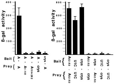
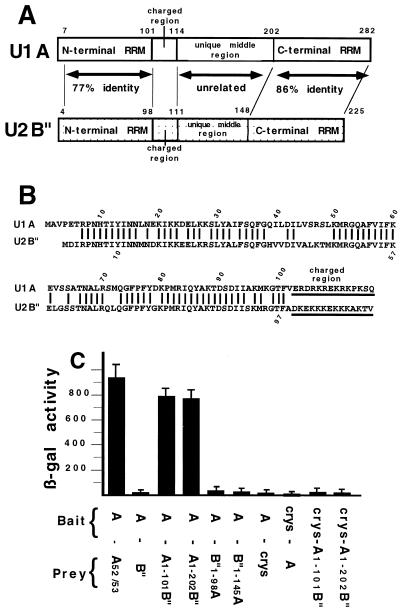
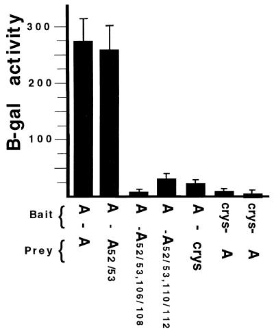
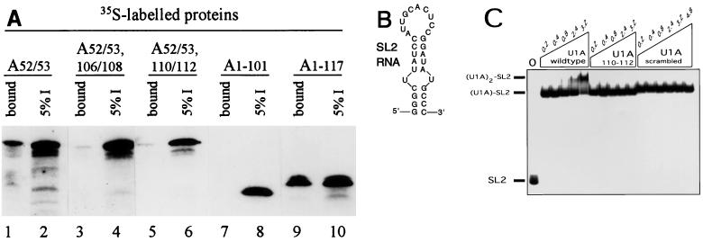
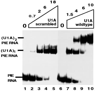
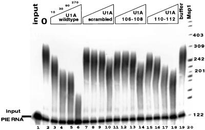
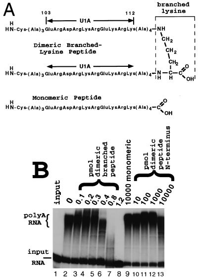
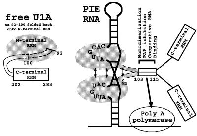
It was previously shown that the human U1A protein, one of three U1 small nuclear ribonucleoprotein-specific proteins, autoregulates its own production by binding to and inhibiting the polyadenylation of its own pre-mRNA. The U1A autoregulatory complex requires two molecules of U1A protein to cooperatively bind a 50-nucleotide polyadenylation-inhibitory element (PIE) RNA located in the U1A 3' untranslated region. Based on both biochemical and nuclear magnetic resonance structural data, it was predicted that protein-protein interactions between the N-terminal regions (amino acids [aa] 1 to 115) of the two U1A proteins would form the basis for cooperative binding to PIE RNA and for inhibition of polyadenylation. In this study, we not only experimentally confirmed these predictions but discovered some unexpected features of how the U1A autoregulatory complex functions. We found that the U1A protein homodimerizes in the yeast two-hybrid system even when its ability to bind RNA is incapacitated. U1A dimerization requires two separate regions, both located in the N-terminal 115 residues. Using both coselection and gel mobility shift assays, U1A dimerization was also observed in vitro and found to depend on the same two regions that were found in vivo. Mutation of the second homodimerization region (aa 103 to 115) also resulted in loss of inhibition of polyadenylation and loss of cooperative binding of two U1A protein molecules to PIE RNA. This same mutation had no effect on the binding of one U1A protein molecule to PIE RNA. A peptide containing two copies of aa 103 to 115 is a potent inhibitor of polyadenylation. Based on these data, a model of the U1A autoregulatory complex is presented.


Figures

FIG. 1
FIG. 1
Two-hybrid analysis of U1A. The two panels represent two independent experiments. Each histogram represents the mean value of three independent transformants, and standard deviations are indicated. The y axis is in β-gal units. Indicated below each histogram are the identities of the fusion proteins expressed in the cells and whether the fusion proteins come from the bait or the prey plasmid. (Left) Analysis of wild-type and deletion mutant forms of U1A. (Right) Analysis of U1A(52/53) mutant proteins. U1C is the human U1 snRNP-specific U1C protein, and crys is the αBcrystallin protein.
FIG. 2
FIG. 2
Two-hybrid analysis of U1A/U2B" chimeras. Structural features of U1A (31) and U2B" (13) and results of yeast two-hybrid analysis of chimeras of U1A and U2B". (A) The open box represents the U1A protein, and the speckled box represents the U2B" protein. Both the U1A and the U2B" proteins contain two RRMs separated by an unrelated middle region. The charged region which is found in both proteins is also indicated. (B) Sequence alignment of the N-terminal portion of the human U1A (above) and U2B" (below) proteins. Only identical residues are indicated by a line. The position of the charged region is also indicated. (C) Results of two-hybrid analysis of the chimeric U1A/U2B" proteins. Chimera U1A(1-101)-B" is aa 1 to 101 of U1A and aa 101 to 225 of U2B", chimera U1A(1-202)-B" is aa 1 to 202 of U1A and aa 148 to 225 of U2B", chimera B"(1-98)-U1A is aa 1 to 98 of U2B" and aa 104 to 282 of U1A, and chimera B" (1-145)-U1A is aa 1 to 145 of U2B" and aa 205 to 282 of U1A. The features of the histograms are as described in the legend to Fig. 1.
FIG. 3
FIG. 3
Two-hybrid analysis of U1As mutated in the charged domain. The features of the histograms are as described in the legend to Fig. 1.
FIG. 4
FIG. 4
U1A homodimerizes in vitro using the same domains as in vivo. (A) Results of histidine tag coselection experiments using an anti-his polyclonal antibody coupled to agarose beads. A 13-pmol (400-ng) sample of recombinant U1A(his) (histidine tagged) was incubated with 35S-labeled mutant U1A proteins (as indicated above the autoradiogram); this was followed by coselection with anti-his antibody coupled to agarose beads. After extensive washing of the beads, the bound proteins were eluted in SDS buffer and analyzed by SDS-PAGE and autoradiography. The even-numbered lanes represent 5% of the total input of 35S-labeled protein, and the odd-numbered lanes represent 50% of the bound proteins that were eluted. (B) Sequence of the SL2 RNA oligonucleotide used in the gel shift experiments in panel C. (C) EMSA of wild-type and mutant U1A proteins binding to the SL2 RNA oligonucleotide. Each lane contains 1 nM 32P-end-labeled SL2 RNA oligonucleotide. Lanes 2 to 17 contain, in addition, increasing micromolar concentrations of wild-type U1A (lanes 2 to 6), U1A(110/112) mutant protein (lanes 7 to 11), or U1A(scrambled) mutant protein (lanes 12 to 17), as indicated above the autoradiogram. On the left are indicated the positions of the unbound SL2 RNA, the (U1A)1-SL2 RNA complex, and the (U1A)2-SL2 RNA complex.
FIG. 5
FIG. 5
The U1A(scrambled) mutant protein has lost cooperative binding to PIE RNA. EMSA of wild-type U1A and U1A(scrambled) mutant protein binding to 32P-labeled PIE RNA. Each lane contains 0.5 nM 32P-labeled PIE RNA. Lanes 2 to 5 and 7 to 10 contain, in addition, increasing nanomolar concentrations of recombinant wild-type U1A or U1A(scrambled), as indicated above the autoradiogram. On the left are indicated the positions of PIE RNA, the (U1A)1-PIE RNA complex, and the (U1A)2-PIE RNA complex. Note that the amounts of U1A used here are ∼1,000-fold less than those used for Fig. 4C.
FIG. 6
FIG. 6
The U1A dimerization domain (aa 103 to 115) is also needed for PAP inhibition. Shown is the inhibition of PAP by either wild-type U1A (lanes 3 to 6), U1A(scrambled) (lanes 7 to 10), U1A(106/108) (lanes 11 to 14), or U1A(110/112) (lanes 15 to 18). Above the autoradiogram is indicated the nanomolar concentration of U1A present in each lane. Polyadenylated RNAs were separated on a denaturing polyacrylamide gel. Each lane contains polyadenylation buffers and 1 nM 32P-labeled PIE RNA incubated either in the absence (lane 1) or in the presence (lanes 2 to 19) of 5 nM recombinant bovine PAP. Lane 19 contains U1A storage buffer in place of U1A protein as a control. Lane 20 is a 32P-end-labeled MspI digest of pBR322, and the sizes of the bands are indicated in nucleotides.
FIG. 7
FIG. 7
Two copies of the aa 103 to 115 dimerization domain are sufficient to inhibit PAP. (A) Schematic of the structure of the monomeric peptide and the dimeric branched-lysine peptide used in the polyadenylation assay shown in panel B. Note that the monomeric peptide has an N-terminal cysteine to allow homodimerization by disulfide bond formation under reducing conditions. (B) The polyadenylation assay is as described in the legend to Fig. 6, except that the chemically synthesized peptides shown in panel A were used in place of U1A. Lanes 1 and 2 are as in Fig. 6. Lanes 2 to 13 contain 5 nM recombinant PAP. Lanes 3 to 8 contain increasing amounts of the dimeric branched-lysine peptide. Lane 9 contains 10,000 pmol of the monomeric peptide. Lanes 10 to 13 contain increasing amounts of the homodimeric peptide covalently linked N terminus to N terminus via cysteine disulfide bonds. The amount of peptide used in each lane is indicated in picomoles above the autoradiogram.
FIG. 8
FIG. 8
The U1A dimerization model incorporates results from a number of publications, including this one, as described in the text. Shown is a schematic of the structure of free U1A protein in order to illustrate that aa 92 to 102 are in a different conformation when the protein is RNA bound. Also shown are two U1A proteins in complex with PIE RNA. The region containing amino acids 92 to 115 of both U1A proteins is involved in protein-protein interactions, as indicated by the arrows. The dimerization of aa 103 to 113 is sufficient to create a surface that promotes both cooperative binding to PIE RNA and binding to and inhibition of vertebrate PAP. Note that the two copies of aa 92 to 115 are deliberately aligned in parallel to each other. In addition, stabilizing protein-protein interactions are present within the N-terminal 100 residues, as indicated by arrows.

References

    1. Allain F H-T, Gubser C C, Howe P W A, Nagai K, Neuhaus D, Varani G. Specificity of ribonucleoprotein interaction determined by RNA folding during complex formation. Nature. 1996;380:646–650. - PubMed
    1. Avis J M, Allain F H-T, Howe P, Varani G, Nagai K, Neuhaus D. Solution structure of the N-terminal RNP domain of U1A protein: the role of the C-terminal residues in structure stability and RNA binding. J Mol Biol. 1996;257:398–411. - PubMed
    1. Boelens W, Scherly D, Jansen E J R, Kolen K, Mattaj I W, van Venrooij W J. Analysis of in vitro binding of U1-A protein mutants to U1 snRNA. Nucleic Acids Res. 1991;19:4611–4618. - PMC - PubMed
    1. Boelens W C, Jansen E J R, van Venrooij W J, Stripecke R, Mattaj I W, Gunderson S I. The human U1 snRNP-specific U1A protein inhibits polyadenylation of its own pre-mRNA. Cell. 1993;72:881–892. - PubMed
    1. Boelens W C, Croes Y, de Ruwe M, de Reu L, De Jong W W. Negative charges in the C-terminal domain stabilize the alpha-βcrystallin complex. J Biol Chem. 1998;273:28085–28090. - PubMed

Publication types

LinkOut - more resources