. 2000 Mar;20(6):2218-27.
doi: 10.1128/MCB.20.6.2218-2227.2000.

Two independent signaling pathways mediate the antiapoptotic action of macrophage-stimulating protein on epithelial cells

Affiliations

Two independent signaling pathways mediate the antiapoptotic action of macrophage-stimulating protein on epithelial cells

A Danilkovitch et al. Mol Cell Biol. 2000 Mar.

Abstract

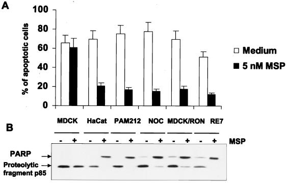
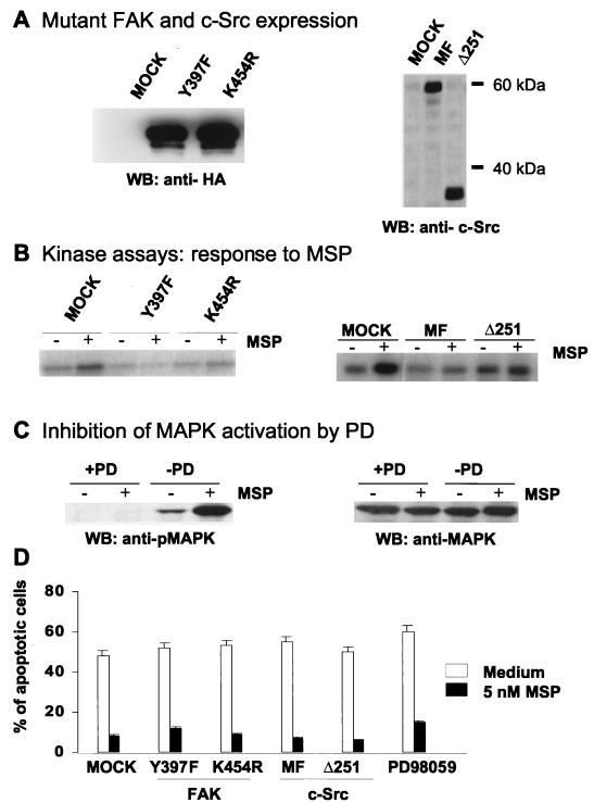
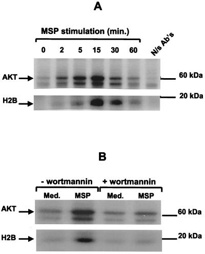
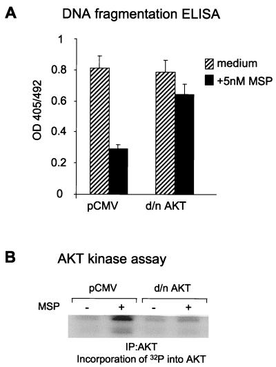
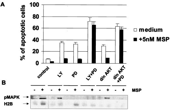
In addition to its effects on macrophage function, macrophage-stimulating protein (MSP) is a growth and motility factor for epithelial cells. The growth and survival of epithelial cells generally require two signals, one generated by interaction with extracellular matrix via integrins, the other initiated by a growth factor. Therefore we investigated the effect of MSP on epithelial cell survival. Survival of epithelial cells cultured overnight in serum-free medium was promoted by adhesion, which activated both the phosphatidylinositol 3'-kinase (PI3-K)/AKT and mitogen-activated protein kinase (MAPK) pathways, operating independently of one another. The number of apoptotic cells resulting from inhibition of either pathway alone was approximately doubled by simultaneous inhibition of both pathways. This shows that each pathway made a partial contribution to the prevention of apoptosis. In the presence of an inhibitor of either pathway, MSP increased the activity of the other pathway so that the single uninhibited pathway alone was sufficient to prevent apoptosis. In contrast to the results with adherent cells, although MSP also prevented apoptosis of cells in suspension (anoikis), its effect was mediated only by the PI3-K/AKT pathway. Despite activation of MAPK by MSP, anoikis was not prevented in suspended cells with a blocked PI3-K/AKT pathway. Thus, activation of MAPK alone is not sufficient to mediate MSP antiapoptotic effects. Cell adhesion generates an additional signal, which is essential for MSP to use MAPK in an antiapoptotic pathway. This may involve translocation of MSP-activated MAPK from the cytoplasm into the nucleus, which occurs only in adherent cells. Our results suggest that there is cross talk between cell matrix adhesion and growth factors in the regulation of cell survival via the MAPK pathway. Growth factors induce MAPK activation, and adhesion mediates MAPK translocation from the cytoplasm into the nucleus.


Figures

FIG. 1
FIG. 1
MSP prevents anoikis in RE7 cells. For induction of anoikis, epithelial cells from regular petri dishes were transferred to poly-Hema-coated dishes. Poly-Hema prevents the attachment of cells to plastic, and nonattached cells undergo apoptosis, which was quantified by TUNEL staining after overnight incubation in serum-free medium (A and B) or in the presence of 5 nM MSP (C and D). Cells attached to uncoated petri dishes in serum-free medium were also evaluated (E and F). Left panels, 4′,6-diamidino-2-phenylindole (DAPI)-stained blue nuclei demonstrating the total number of cells in the field; right panels, fluorescein isothiocyanate-stained apoptotic cells in the same field.
FIG. 2
FIG. 2
MSP prevents anoikis in various RON-expressing keratinocyte and epithelial cell lines. Anoikis was induced as described in the legend to Fig. 1. (A) The number of apoptotic cells was determined by TUNEL staining. Each experimental point represents the mean percentage (+ standard error of the mean) of apoptotic cells for three independent experiments. (B) Detection of apoptosis by immunoblot analysis of PARP cleavage in total cell lysates.
FIG. 3
FIG. 3
Inhibition of MSP-induced FAK, c-Src, or MAPK activation does not prevent the antianoikis effect of MSP. (A) RE7 cells were transiently transfected with dominant-negative FAK Y397F or K454R or c-Src MF or Δ251 or empty vector (mock). Lysates (15 μl/lane) from transfected and MACS4-selected cells were analyzed by SDS-PAGE followed by Western blotting with anti-HA antibodies for detection of FAK expression (left) and with anti-c-Src antibodies for detection of c-Src expression (right) (B) A suspension of transiently transfected RE7 cells (2 × 10⁶/ml) was stimulated with 5 nM MSP for 10 min. FAK and c-Src kinases were then immunoprecipitated from cell lysates with anti-FAK or anti-c-Src antibodies. The kinase activities of FAK and c-Src were measured with [32P]ATP as the capacity of the FAK IP to autophosphorylate FAK (left) or the c-Src IP to phosphorylate enolase (right), detected by SDS-PAGE and autoradiography. (C) A suspension of RE7 cells was pretreated with 50 μM MEK inhibitor PD98059 for 15 min, and then the cells were stimulated with 5 nM MSP for an additional 15 min. Activation of MAPK was determined by Western blotting with anti-phosphoMAPK (pMAPK) (left) or anti-MAPK (right) after SDS-PAGE of total cell lysates. (D) Anoikis was induced in mutant FAK or c-Src-transfected RE7 cells as described in the legend to Fig. 1. After overnight incubation in medium with or without 5 nM MSP, the apoptotic cells were quantified by TUNEL staining. Each experimental point represents the mean percentage (+ standard error of the mean) of apoptotic cells for three independent experiments.
FIG. 4
FIG. 4
MSP stimulates AKT enzymatic activity by a PI3-K-dependent mechanism. (A) After stimulation of RE7 cells with 5 nM MSP, the cells were lysed at the indicated times. AKT was immunoprecipitated and tested for kinase activity by incubation with [32P]ATP and exogenous substrate histone H2B. Incorporation of 32P into AKT (top) and histone H2B (bottom) was detected by SDS-PAGE and autoradiography. N/s Ab's, antibodies isolated from nonimmune rabbit serum. (B) RE7 cells were pretreated with 100 nM WTM for 15 min and then stimulated with 5 nM MSP for an additional 15 min. AKT kinase activity was detected as described for panel A.
FIG. 5
FIG. 5
Dominant-negative AKT blocks the antianoikis effect of MSP. (A) RE7 cells were transiently transfected with kinase-dead AKT (d/n AKT) or with the pCMV vector as a control. Anoikis was induced as described in the legend to Fig. 1, and DNA fragmentation in apoptotic cells was quantified by cell death ELISA. Three independent experiments were performed. The results of a representative experiment are shown. The error bars indicate the standard deviations for triplicate ELISA wells. (B) Expression of dominant-negative AKT blocks MSP-induced endogenous AKT enzymatic activity. RE7 cells, transiently transfected with the empty vector (pCMV) or dominant-negative AKT cDNA, were stimulated with 5 nM MSP for 15 min. The cells were then lysed, and AKT was immunoprecipitated and tested for kinase activity in vitro. The kinase assay was performed as described in the legend to Fig. 4A. Incorporation of 32P into AKT was detected by SDS-PAGE and autoradiography.
FIG. 6
FIG. 6
MSP rescues epithelial cells from serum deprivation-induced apoptosis via PI3-K/AKT and MAPK pathways. (A) The effect of MSP on RE7 cells or RE7 cells with transiently expressed dominant-negative AKT was studied in serum-free medium in the presence of inhibitor LY294002 or PD98059 or both combined. After 24 h of incubation, the percentage of apoptotic cells was determined by TUNEL staining. Each experimental point represents the mean percentage (+ standard deviation) of apoptotic cells in three independent experiments. (B) AKT kinase activity was determined as phosphorylation of histone H2B in vitro as described in the legend to Fig. 3. MAPK activity was determined in total cell lysates by Western blotting with antibodies specific for phosphorylated MAPK.
FIG. 7
FIG. 7
Adhesion promotes translocation of MSP-activated MAPK from the cytoplasm into the nucleus. Starved RE7 epithelial cells were stimulated with 5 nM MSP for 15 min in suspension or on coverslips, and then translocation of MAPK was detected by indirect immunofluorescence analysis (A) or by Western blotting of cytoplasmic and nuclear cell extracts (B) with antibodies to phosphoMAPK (pMAPK) (top) or total MAPK (bottom).

References

    1. Alessi D R, Cohen P. Mechanism of activation and function of protein kinase B. Curr Opin Genet Dev. 1998;8:55–62. - PubMed
    1. Alessi D R, Cuenda A, Cohen P, Dudley D T, Saltiel A R. PD 098059 is a specific inhibitor of the activation of mitogen-activated protein kinase kinase in vitro and in vivo. J Biol Chem. 1995;270:27489–27494. - PubMed
    1. Aplin A E, Juliano R L. Integrin and cytoskeletal regulation of growth factor signaling to the MAP kinase pathway. J Cell Sci. 1999;112:695–706. - PubMed
    1. Assoian R K, Zhu X. Cell anchorage and the cytoskeleton as partners in growth factor dependent cell cycle progression. Curr Opin Cell Biol. 1997;9:93–98. - PubMed
    1. Berra E, Diaz-Meco M T, Moscat J. The activation of p38 and apoptosis by the inhibition of Erk is antagonized by the phosphoinositide 3-kinase/Akt pathway. J Biol Chem. 1998;273:10792–10797. . (Erratum, 273:16630.) - PubMed