. 2000 Mar;20(6):2239-47.
doi: 10.1128/MCB.20.6.2239-2247.2000.

Stimulus-specific assembly of enhancer complexes on the tumor necrosis factor alpha gene promoter

Affiliations

Stimulus-specific assembly of enhancer complexes on the tumor necrosis factor alpha gene promoter

J V Falvo et al. Mol Cell Biol. 2000 Mar.

Abstract

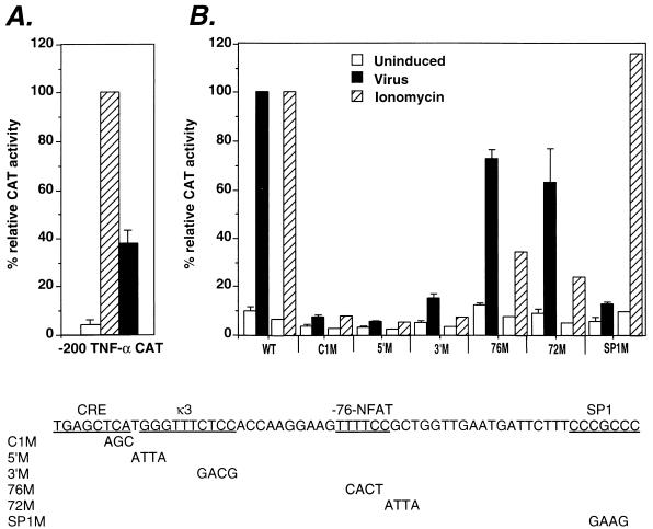
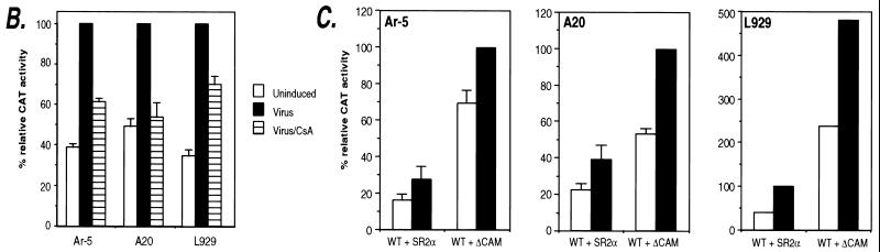
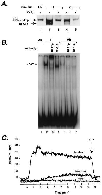
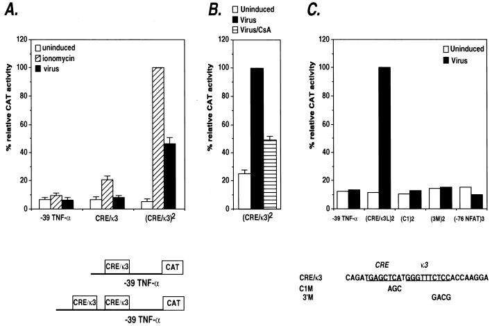
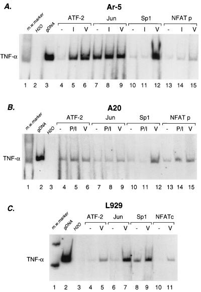
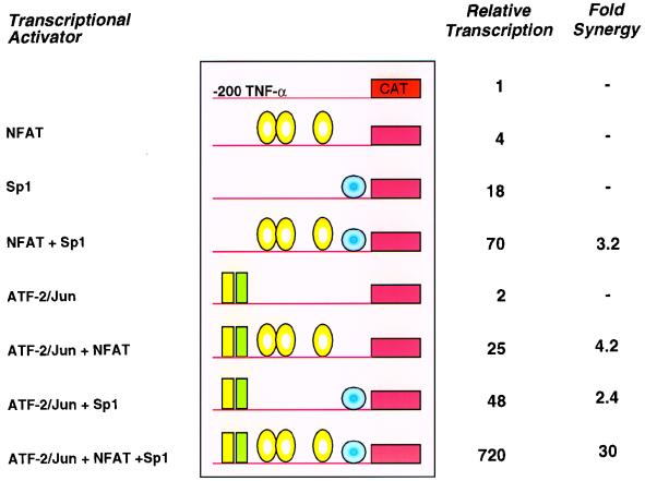
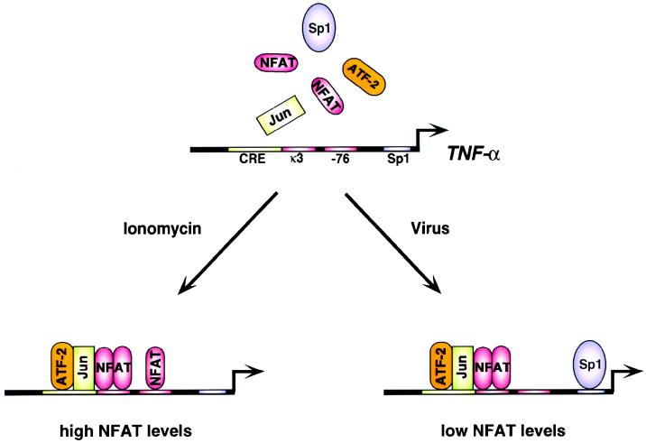
The human tumor necrosis factor alpha (TNF-alpha) gene is rapidly activated in response to multiple signals of stress and inflammation. We have identified transcription factors present in the TNF-alpha enhancer complex in vivo following ionophore stimulation (ATF-2/Jun and NFAT) and virus infection (ATF-2/Jun, NFAT, and Sp1), demonstrating a novel role for NFAT and Sp1 in virus induction of gene expression. We show that virus infection results in calcium flux and calcineurin-dependent NFAT dephosphorylation; however, relatively lower levels of NFAT are present in the nucleus following virus infection as compared to ionophore stimulation. Strikingly, Sp1 functionally synergizes with NFAT and ATF-2/c-jun in the activation of TNF-alpha gene transcription and selectively associates with the TNF-alpha promoter upon virus infection but not upon ionophore stimulation in vivo. We conclude that the specificity of TNF-alpha transcriptional activation is achieved through the assembly of stimulus-specific enhancer complexes and through synergistic interactions among the distinct activators within these enhancer complexes.


Figures

FIG. 1
FIG. 1
Virus induction of TNF-α gene requires the CRE/κ3 and SP1 sites. (A) Relative activation levels of the −200 TNF-α–CAT fusion construct by ionophore and virus. The Ar-5 T-cell clone was transfected with the −200 TNF-α promoter–CAT reporter construct. Cells were mock stimulated (Uninduced) or stimulated with ionomycin (Ionomycin) or Sendai virus (Virus). CAT assays were performed, and the percent conversion of [14C]chloramphenicol to its acetylated forms was quantified. Within individual experiments, the results were normalized to the −200 TNF-α–CAT ionomycin-induced level (100%) and then averaged and plotted in the displayed histogram. Standard deviation is represented by the error bars. All cells were cotransfected with the cytomegalovirus β-Gal plasmid, and β-Gal assays were performed to control for transfection efficiency. The figure shows the results of three independent experiments. (B) Relative activities of human TNF-α–CAT fusion constructs containing mutations in the CRE, κ3-NFAT, −76-NFAT, or SP1 site in virus-stimulated Ar-5 T cells. Ar-5 T cells were transfected with the wild-type −200 TNF-α promoter–CAT reporter (WT) and mutated −200 TNF-α promoter–CAT reporter genes (C1M, 5′M, 3′M, 76M, 72M, SP1M), and CAT assays were performed and quantified as described above. The figure shows the results of three independent experiments. Previously published results showing the effect of these mutations upon ionomycin induction of the gene (13, 44, 45) are included in the figure for comparison. Each ionomycin set is independently normalized to the −200 WT ionomycin-induced level (100%) on the same graph as the virus set, which is independently normalized to the −200 WT virus-induced level (100%).
FIG. 2
FIG. 2
Calcineurin is involved in virus-inducible TNF-α gene induction. (A) Induction of TNF-α mRNA by ionophore and virus in Ar-5 T cells. An autoradiogram of an RNase protection assay mapping TNF-α and γ-actin mRNAs is shown. Ar-5 cells were stimulated with ionomycin (I) for 30 min or with Sendai virus (vir) for 2 h in the presence or absence of CsA. The γ-actin probe was made to have a specific activity that was one-fifth the specific activity of the mouse TNF-α probe. (B) CsA inhibits virus induction of TNF-α CAT reporter activity. Ar-5 T cells, A20 B cells, or L929 fibroblasts were transfected with the −200 TNF-α–CAT reporter gene. Twenty-four hours after transfection, the cells were mock induced (Uninduced) or stimulated with Sendai virus (Virus) in the presence or absence of CsA as indicated in the figure. CAT assays were performed and quantified as described in the legend to Fig. 1. The figure shows the results of three independent experiments. (C) Calcineurin augments virus induction of TNF-α. Ar-5 T cells, A20 B cells, or L929 fibroblasts were transfected with the −200 TNF-α–CAT reporter gene and a plasmid that constitutively expresses the catalytic subunit of calcineurin (ΔCAM) or a control plasmid (SR2α) as indicated in the figure. Twenty-four hours after transfection, the cells were either mock induced (Uninduced) or stimulated with Sendai virus (Virus) as described for panel B. CAT assays were performed and quantified as described in the legend to Fig. 1. The figure shows the results of three independent experiments for Ar-5 and A20 cells and a representative experiment for L929 cells.
FIG. 2
FIG. 2
Calcineurin is involved in virus-inducible TNF-α gene induction. (A) Induction of TNF-α mRNA by ionophore and virus in Ar-5 T cells. An autoradiogram of an RNase protection assay mapping TNF-α and γ-actin mRNAs is shown. Ar-5 cells were stimulated with ionomycin (I) for 30 min or with Sendai virus (vir) for 2 h in the presence or absence of CsA. The γ-actin probe was made to have a specific activity that was one-fifth the specific activity of the mouse TNF-α probe. (B) CsA inhibits virus induction of TNF-α CAT reporter activity. Ar-5 T cells, A20 B cells, or L929 fibroblasts were transfected with the −200 TNF-α–CAT reporter gene. Twenty-four hours after transfection, the cells were mock induced (Uninduced) or stimulated with Sendai virus (Virus) in the presence or absence of CsA as indicated in the figure. CAT assays were performed and quantified as described in the legend to Fig. 1. The figure shows the results of three independent experiments. (C) Calcineurin augments virus induction of TNF-α. Ar-5 T cells, A20 B cells, or L929 fibroblasts were transfected with the −200 TNF-α–CAT reporter gene and a plasmid that constitutively expresses the catalytic subunit of calcineurin (ΔCAM) or a control plasmid (SR2α) as indicated in the figure. Twenty-four hours after transfection, the cells were either mock induced (Uninduced) or stimulated with Sendai virus (Virus) as described for panel B. CAT assays were performed and quantified as described in the legend to Fig. 1. The figure shows the results of three independent experiments for Ar-5 and A20 cells and a representative experiment for L929 cells.
FIG. 3
FIG. 3
Virus infection causes NFAT dephosphorylation and an increase in intracellular calcium levels. (A) Infection of Ar-5 T cells by Sendai virus causes dephosphorylation of NFATp. Nuclear extracts were prepared from Ar-5 T cells stimulated with ionomycin (I) for 30 min or with Sendai virus (Vir) for 2 h, in the presence or absence of CsA as indicated in the figure. Lysates were analyzed by sodium dodecyl sulfate–6% polyacrylamide gel electrophoresis followed by Western blot analysis with the anti-NFATp (67.1) antibody. After ionomycin or virus stimulation of the cells, phosphorylated NFATp (P-NFATp) is dephosphorylated (NFATp) and represented as a size shift in the gel. (B) Virus-inducible NFATp/c binds to the −76-NFAT site. An EMSA using nuclear extracts from unstimulated Ar-5 cells (UN) or cells stimulated with ionomycin (I) for 30 min or with Sendai virus (Vir) for 2 h as indicated in the figure was performed. An ionomycin- and virus-inducible complex binds to the oligonucleotide probe spanning positions −85 to −59 containing the −76-NFAT site. Antibodies to NFATp and to NFATc specifically react with the ionomycin- and virus-inducible complex, since they do not react with complexes bound to an Sp1 oligonucleotide probe (47). (C) Infection of Ar-5 T cells by Sendai virus causes an increase in intracellular calcium levels. Cells were exposed to either Sendai virus or ionomycin or a control carrier (allantoic fluid in which the Sendai virus is prepared) at 0 min. Extracellular calcium was later buffered to 0 mM by the addition of 4 mM EGTA (arrow).
FIG. 4
FIG. 4
Inducer-specific regulation of the CRE/κ3 composite element by ionophore and virus. (A) Activation of synthetic TNF-α promoters containing the CRE/κ3 multimers by ionomycin or virus in Ar-5 T cells. Ar-5 cells were transfected with the truncated −39 TNF-α–CAT construct containing either one or two copies of the κ3(L) site. Twenty-four hours later, the cells were mock stimulated (uninduced) or stimulated with ionomycin (ionomycin) or Sendai virus (virus). Within the individual experiments displayed, the results were normalized to the induced level (100%) of the wild-type (CRE/κ3)2 multimer construct and then averaged and plotted in the displayed histogram. CAT assays were performed and quantified as described in the legend to Fig. 1. The figure shows the results of three independent experiments. (B) CsA inhibits virus activation of the CRE/κ3 site. Ar-5 cells were transfected with the κ3(L)2 synthetic promoter construct, stimulated with virus in the presence or absence of CsA, and analyzed and quantified as described in the legend to Fig. 1. The figure shows the results of three independent experiments. (C) Activation of synthetic TNF-α promoters containing CRE/κ3 or −76-NFAT multimers in Ar-5 T cells. Ar-5 cells were transfected with the truncated −39 TNF-α–CAT construct containing either two copies of the wild-type CRE/κ3 site or two copies of the CRE/κ3 site containing the 3′ mutation or the C1 mutation or with three copies of the −76-NFAT site as indicated in the figure. Twenty-four hours later, the cells were mock stimulated (Uninduced) or stimulated with Sendai virus (Virus) as indicated. The figure shows a representative experiment. The results were normalized to the induced level (100%) of the wild-type (CRE/κ3)2 multimer construct and then averaged and plotted in the displayed histogram.
FIG. 5
FIG. 5
Association of specific transcriptional activator proteins with the TNF-α promoter in vivo upon ionophore stimulation and virus induction. Ar-5 T cells (A), A20 B cells (B), and L929 fibroblasts (C) were unstimulated (−) or stimulated with Sendai virus (V), ionomycin (I), or PMA plus ionomycin (P/I) for 3 h as indicated. The cells were then treated with formaldehyde to cross-link protein to DNA, and chromatin was isolated and purified and immunoprecipitated with the indicated antibodies as described previously (34, 50). After reversal of the cross-links, the DNA was amplified by PCR with primers to TNF-α (−210 to +36) (PCRs, 20 cycles of 1 min each at 94, 70, and 72°C). A range of PCR cycles was performed to ensure that the products visualized were in the linear range of amplification. Control amplification using genomic DNA (gDNA) and water (H20) are shown along with a 123-bp marker (Life Technologies; m.w. marker). The high levels of constitutive binding of c-jun to the TNF-α promoter in Ar-5 cells in the chromatin immunoprecipitation assay are consistent with studies demonstrating that c-jun homodimers can bind the TNF-α CRE (44). Although low relative increases in levels of amplified DNA were observed in the NFATp immunoprecipitations, the antibody used was the only one of those tested that was suitable for the chromatin immunoprecipitation assay, and results with Ar-5 and A20 cells were consistent.
FIG. 6
FIG. 6
Sp1 synergizes with NFATp and ATF-2/c-jun. Drosophila SL2 cells were cotransfected with NFATp S>A (2.4 μg), Sp1 (100 ng), and ATF-2/c-jun (500 ng) in the combinations indicated along with Hsp–β-Gal (500 ng) and the −200 TNF-α–CAT reporter (500 ng). CAT activity normalized for β-Gal expression is shown for a representative experiment. The fold synergy of activation (i.e., fold activation greater than the additive effect of the individual activators) is noted. The experiment shown is representative of two independent experiments. The experiment testing the synergy between NFATp and Sp1 was performed five independent times. We note that increasing amounts (0.1 to 2.4 μg) of NFATp S>A cotransfected into SL2 cells with the TNF-α–CAT reporter gene resulted in up to a fourfold increase of TNF-α-driven CAT activity (31).
FIG. 7
FIG. 7
Inducer-specific assembly of distinct enhancer complexes on the TNF-α gene promoter. The cis-acting TNF-α promoter elements, transcription factors, and coactivators involved in the inducer-specific regulation of TNF-α by ionophore and virus in T cells are summarized schematically. The sites that are critical for activation of the promoter are indicated. The transcription factors which bind to the TNF-α promoter in vivo (NFAT, ATF-2/c-jun, and Sp1) following the indicated stimuli are shown.

References

    1. Aderka D, Holtmann H, Toker L, Hahn T, Wallach D. Tumor necrosis factor induction by Sendai virus. J Immunol. 1986;136:2938–2942. - PubMed
    1. Aggarwal B B, Puri R K. Human cytokines: their role in disease and therapy. Cambridge, Mass: Blackwell Science; 1995.
    1. Beals C R, Clipstone N A, Ho S N, Crabtree G R. Nuclear localization of NF-ATc by a calcineurin-dependent, cyclosporin-sensitive intramolecular interaction. Genes Dev. 1997;11:824–834. - PubMed
    1. Boussiotis V A, Nadler L M, Strominger J L, Goldfeld A E. Tumor necrosis factor α is an autocrine growth factor for normal human B cells. Proc Natl Acad Sci USA. 1994;91:7007–7011. - PMC - PubMed
    1. Brinkman B M N, Telliez J-B, Schievella A R, Lin L-L, Goldfeld A E. Engagement of TNF receptor 1 leads to ATF-2 and p38 MAP kinase-dependent TNF-α gene expression. J Biol Chem. 1999;274:30882–30886. - PubMed

Publication types

MeSH terms

LinkOut - more resources