. 2000 Feb 29;97(5):2253-8.
doi: 10.1073/pnas.040565597.

Marking synaptic activity in dendritic spines with a calpain substrate exhibiting fluorescence resonance energy transfer

Affiliations

Marking synaptic activity in dendritic spines with a calpain substrate exhibiting fluorescence resonance energy transfer

P W Vanderklish et al. Proc Natl Acad Sci U S A. .

Abstract

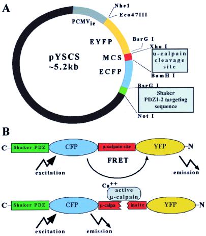
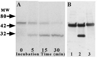
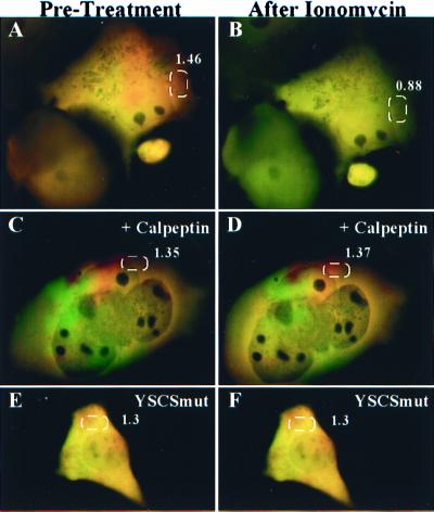
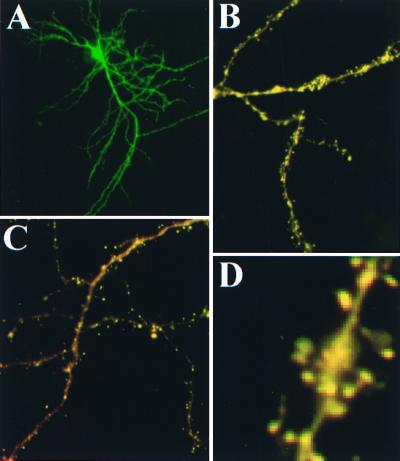
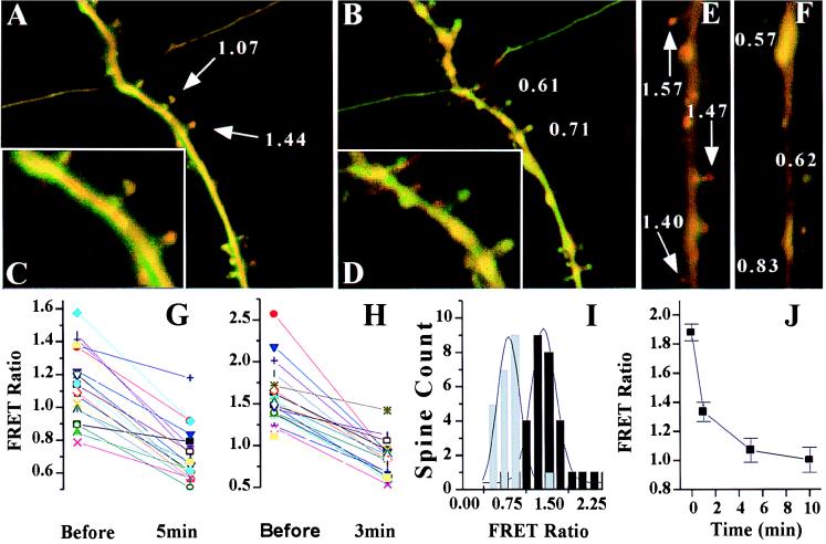
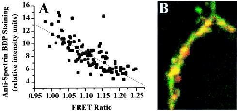
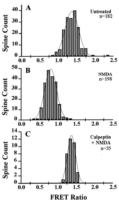
Excitatory synaptic activity can evoke transient and substantial elevations of postsynaptic calcium. Downstream effects of elevated calcium include the activation of the calcium-dependent protease calpain. We have developed a reagent that identifies dendritic spines in which calpain has been activated. A fusion protein was expressed that contained enhanced yellow and enhanced cyan fluorescent protein (EYFP and ECFP, respectively) linked by a peptide that included the micro-calpain cleavage site from alpha-spectrin. A PDZ-binding site fused to ECFP anchored this protein to postsynaptic densities. The fusion protein exhibited fluorescence resonance energy transfer (FRET), and diminution of FRET by proteolysis was used to localize calpain activity in situ by fluorescence microscopy. Incubation of the fusion protein with calpain in the presence of calcium resulted in the separation of EYFP and ECFP into monomeric fluorophores. In transiently transfected cell lines and dissociated hippocampal neurons, FRET was diminished by raising intracellular calcium levels with an ionophore or with glutamatergic agonists. Calpain inhibitors blocked these changes. Under control conditions, FRET levels in different dendritic spines of cultured neurons and in hippocampal slices were heterogeneous but showed robust decreases upon treatment with glutamatergic agonists. Immunostaining of cultured neurons with antibodies to a spectrin epitope produced by calpain-mediated digestion revealed an inverse correlation between the amount of FRET present at postsynaptic elements and the concentration of spectrin breakdown products. These results suggest that the FRET methodology identifies sites of synaptically induced calpain activity and that it may be useful in analyzing synapses undergoing changes in efficacy.


Figures

Figure 1
Figure 1
Design of a calpain-sensitive marker, pYSCS. (A) Using the pEGFP-C3 vector (CLONTECH), EYFP and ECFP were fused in-frame with an intervening sequence from the μ-calpain cleavage site in α-spectrin. A PDZ cognate sequence that binds to PSD95 was added at the 3′ end to facilitate retention of the expressed protein at postsynaptic sites. (B) Expression of pYSCS yielded a fusion protein from which FRET could be visualized in situ after excitation of the donor ECFP fluorophore. Separation of ECFP and EYFP by μ-calpain abolishes FRET, resulting in decreased EYFP fluorescence and a concomitant increase of ECFP fluorescence.
Figure 2
Figure 2
Sensitivity of the YSCS fusion protein to proteolysis by μ-calpain in vitro. Extracts of COS-7 cells transfected with pYSCS were incubated with purified μ-calpain and calcium. (A) A Western blot of samples harvested at the indicated times was probed with an anti-GFP antibody that recognized both the 54-kDa YSCS protein and a 27-kDa band corresponding to the molecular mass of monomeric GFPs (27 kDa). (B) Inclusion of the calcium chelator EGTA (lane 1) or the calpain inhibitor calpeptin (lane 3) to the reaction mixture blocked formation of the breakdown product. Extracts from cells transfected with pYSCSmut showed partial proteolysis of the protein (lane 2).
Figure 3
Figure 3
YSCS protein exhibits FRET, which is sensitive to calcium levels and calpain activity. COS-7 cells transfected with pYSCS were imaged before (A, C, and E) and 10 min after (B, D, and F) treatment with ionomycin and 6 mM CaCl2. In untreated cells (A), addition of ionomycin (B) produced a conversion of YSCS fluorescence from a mixture of FRET-based (red) and donor (green) signals to an almost purely donor-generated signal. Pretreatment with calpeptin (C) 20 min before addition of ionomycin blocked reductions in FRET-based fluorescence initiated by calcium influx (D). Treatment of N2A cells expressing YSCSmut (E) with ionomycin demonstrated only minor changes in the FRET ratio (F). FRET ratio values averaged over highlighted regions of the cells are shown in white text.
Figure 4
Figure 4
Expression of YSCS in neurons. Low-magnification images of YSCS fluorescence after excitation of YFP in a neuron (A) from an organotypic hippocampal culture. Images collected during CFP excitation reveal the occurrence of both donor (green) and FRET-based fluorescence (red) throughout the dendritic processes of neurons in organotypic (B) and dissociated (C) hippocampal preparations. Enlargement of a dendrite in slice culture (D) demonstrates that YSCS is concentrated in spine heads.
Figure 5
Figure 5
Treatment of dissociated hippocampal neurons with glutamatergic agonists leads to a decrease in FRET. (A) A dendrite with numerous spines from a pyramidal cell transfected with pYSCS and imaged live. FRET and ECFP fluorescence are shown in red and green, respectively. FRET ratios for spines indicated by arrows are shown in white text. (B) The same cell imaged 3 min after addition of 100 μM glutamate. Most spines change from a predominantly FRET signal to a greener donor signal and show reduced FRET ratios. Enlargements of selected spines pretreatment (C) and posttreatment (D) with glutamate are displayed. Paired images are shown from experiments before (E) and after (F) treating dissociated neurons with 100 mM NMDA. Plots of FRET ratios of individual spines are shown before and after treatment with glutamate (G) and NMDA (H). Comparable reductions in FRET ratios were observed in most spines, but some exhibited only slight variations from starting FRET ratios. (I) Histograms of binned FRET ratios from spine populations imaged live showed significant (P < 0.0001) reductions after NMDA treatments (black, pretreatment; gray, posttreatment) and after glutamate treatment (data not shown). (J) Dissociated cultures were fixed at different time points after treatment with NMDA, and the FRET ratio was measured. n = 30, 26, 133, and 100 spines for the time points 0, 1, 5, and 10 min, respectively.
Figure 6
Figure 6
Correlation between histochemical and FRET measures of calpain activation in spines. Dissociated hippocampal neurons were transfected with pYSCS and immunolabeled with antibodies specific for calpain-mediated spectrin breakdown products (BDPs). (A) Correlation curve relating the relative intensities of FRET and BDP immunofluorescence in individual spine heads (r = −0.78). (B) Images of stained neurons in which FRET fluorescence is shown in green and anti-BDP staining is shown in red. The majority of the dendrite displays both FRET fluorescence and anti-BDP staining and appears yellow.
Figure 7
Figure 7
Distributions of FRET ratios from dendritic spines in fixed hippocampal slice cultures. (A and B) The effect of NMDA on spine FRET ratio distributions (bins of 100) from CA1 pyramidal neurons in a representative experiment. A 3-min treatment with NMDA caused a significant reduction of spine FRET ratios 30 min later that was blocked by pretreatment with calpeptin (C).

References

    1. Lynch G, Granger R. J Cognit Neurosci. 1992;4:189–199. - PubMed
    1. Roman F S, Chaillan F A, Soumireu-Mourat B. Brain Res. 1993;601:265–272. - PubMed
    1. Maren S. Trends Neurosci. 1999;22:561–567. - PubMed
    1. Malenka R C, Kauer J A, Zucker R S, Nicoll R A. Science. 1988;242:81–84. - PubMed
    1. Lynch G, Larson J, Kelso S, Barrionuevo G, Schottler F. Nature (London) 1983;305:719–721. - PubMed

Publication types

MeSH terms

LinkOut - more resources