

Loss of PTEN facilitates HIF-1-mediated gene expression
Affiliations
- PMID: 10691731
- PMCID: PMC316386
Item in Clipboard
Loss of PTEN facilitates HIF-1-mediated gene expression
Genes Dev.
.
Abstract
In glioblastoma-derived cell lines, PTEN does not significantly alter apoptotic sensitivity or cause complete inhibition of DNA synthesis. However, in these cell lines PTEN regulates hypoxia- and IGF-1-induced angiogenic gene expression by regulating Akt activation of HIF-1 activity. Restoration of wild-type PTEN to glioblastoma cell lines lacking functional PTEN ablates hypoxia and IGF-1 induction of HIF-1-regulated genes. In addition, Akt activation leads to HIF-1alpha stabilization, whereas PTEN attenuates hypoxia-mediated HIF-1alpha stabilization. We propose that loss of PTEN during malignant progression contributes to tumor expansion through the deregulation of Akt activity and HIF-1-regulated gene expression.
Figures
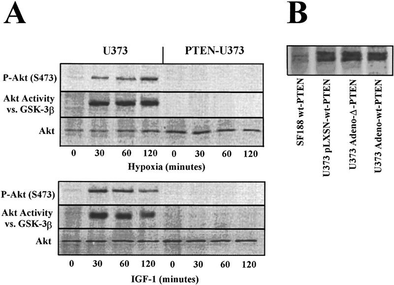
Hypoxia induces Akt activity that is regulated by PTEN. (A) U373 cells were retrovirally infected with wild-type PTEN and subjected to hypoxia or IGF-1 (50 ng/ml) for the indicated times. Cells were harvested and the lysates allocated for immunoblotting using α-Phospho-Akt (S473) (top) α-Akt antibodies or Akt kinase activity using GST–GSK-3β as a substrate. (B) U373 cells were either retrovirally (wt–PTEN) or adenovirally (wt or phosphatase inactive-Δ PTEN) infected. The U373-infected cells or a glioblastoma cell line possessing two wt–PTEN alleles (SF188) as control were immunoprecipitated and immunoblotted with Santa Cruz SC-571 and UBI-ID 07016, respectively.

PTEN overexpression incompletely inhibits DNA synthesis and has minimal effects on apoptosis. U373 cells were infected with adenovirus containing wild-type or phosphatase inactive-Δ PTEN. (A) Forty-eight hours postinfection, serum-starved U373-infected cells were labeled with [3H]-thymidine, treated as indicated for 24 hr, and assayed for [3H]-thymidine incorporation. (B) Forty-eight hours postinfection, U373-infected cells were treated as indicated and assayed for apoptosis.

PTEN regulates the expression of HIF-1-regulated genes under hypoxia. U373 or SF210 cells were retrovirally infected with wild-type PTEN or empty vector control. Thirty-six hours postinfection, U373 and SF210 cells expressing PTEN or empty vector control and parental cells ± 100 nm wortmannin were subjected to 9 hr of hypoxia. mRNA was isolated and analyzed by Northern blot using VEGF, COX-1, PGK-1, PFK, c-fos, and PTEN probes.
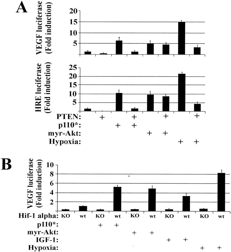
PTEN regulates PI(3)K-induced VEGF expression upstream of Akt in a HIF-1α dependent fashion. (A) U373 cells were cotransfected with either a VEGF–luciferase (top) or a HRE–luciferase (bottom) reporter in combination with either a constitutively active PI(3)K (p110*) or a constitutively active Akt (myr–Akt). Portions of the parental U373 and the p110* and myr–Akt-transfected cells were then retrovirally infected with PTEN. Thirty-six hours postinfection, the cells were treated under oxic or hypoxic conditions for 12 hr, followed by lysis and luciferase activity quantitation. (B) HIF-1α homozygous null and HIF-1α wild-type MEFs were cotransfected with VEGF–luciferase in combination with either a constitutively active PI(3)K (p110*) or a constitutively active Akt (myr–Akt). Portions of these cells were then retrovirally infected with PTEN. Thirty-six hours postinfection, the cells were treated with IGF-1 or exposed to hypoxic conditions for 12 hr followed by lysis and luciferase activity quantitation.

Akt stimulates HIF-1α stabilization. (A) U373 cells were retrovirally infected with wild-type PTEN. Thirty-six hours postinfection, U373 parental cells and PTEN-expressing U373 were subjected to hypoxia for the indicated time. Cells were lysed at the indicated times followed by SDS-PAGE, transfer, and immunoblotting with anti-HIF-1α. (B) Cells were retrovirally infected with myr–Akt–ER or myr–A2–ER. Thirty-six hours postinfection, the cells were subjected to induction by 4-HT for the indicated time and analyzed as in A. (C) U373 cells were serum-deprived and subjected to 1 hr of hypoxia to activate Akt or to UV-C (10 J/m2) for JNK-1 activation for use in immune complex kinase assays. Reactions were performed using 500 ng of either histone H2B, GST–jun, GST–GSK-3β, or HIF-1α as substrates and Akt or JNK-1 immunoprecipitations for in vitro kinase assays. Kinase reactions were subjected to SDS-PAGE, and gels were dried and visualized by PhosphorImaging. (D) Cells were subjected to hypoxia for the indicated times, lysed, immunoprecipitated using anti-HIF-1α, and subjected to SDS-PAGE, transfer, and immunoblot using anti-HIF-1α anti-Akt, or anti-HIF-1β antibodies.
References
-
- Brunet A, Bonni A, Zigmond MJ, Lin MZ, Juo P, Hu LS, Anderson MJ, Arden KC, Blenis J, Greenberg ME. Akt promotes cell survival by phosphorylating and inhibiting a Forkhead transcription factor. Cell. 1999;96:857–868. - PubMed
-
- Campbell SL, Khosravi-Far R, Rossman KL, Clark GJ, Der CJ. Increasing complexity of Ras signaling. Oncogene. 1998;17:1395–1413. - PubMed
-
- Datta SR, Dudek H, Tao X, Masters S, Fu H, Gotoh Y, Greenberg ME. Akt phosphorylation of BAD couples survival signals to the cell-intrinsic death machinery. Cell. 1997;91:231–241. - PubMed
Publication types
MeSH terms
Substances
Grants and funding
LinkOut - more resources
Full Text Sources
Other Literature Sources
Medical
Research Materials
Miscellaneous