

PPD-specific IgG1 antibody subclass upregulate tumour necrosis factor expression in PPD-stimulated monocytes: possible link with disease pathogenesis in tuberculosis
- PMID: 10691916
- PMCID: PMC1905580
- DOI: 10.1046/j.1365-2249.2000.01139.x
PPD-specific IgG1 antibody subclass upregulate tumour necrosis factor expression in PPD-stimulated monocytes: possible link with disease pathogenesis in tuberculosis
Abstract
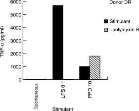
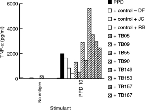
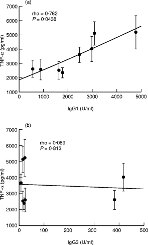
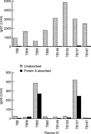
Cachexia is a prominent feature of advanced tuberculosis, in association with increased expression of the monokine tumour necrosis factor (TNF)-alpha. Monocytes, have high affinity receptors (mannose, complement and Fc gamma1 and gamma111) which mediate antigen uptake and subsequent cytokine activation. Several mycobacterial proteins, including PPD, can stimulate TNF-alpha secretion from monocytes. However, the role of various receptors in stimulating or regulating TNF-alpha secretion is still unclear. We have previously shown selective augmentation of opsonic antibodies (IgG1 and IgG3) in tuberculosis patients with advanced pulmonary disease. We now analyse the role of opsonizing antibodies in modulating TNF-alpha expression in antigen stimulated monocytes. PPD was used as the prototypic mycobacterial antigen to stimulate monocytes from PPD skin test negative donors (n = 7) in the presence of plasma from tuberculosis patients (n = 8), containing known amounts of IgG1 and IgG3 anti-PPD antibodies. TNF-alpha secretion was enhanced in the presence of TB plasma (4/8) but not in the presence of control plasma. Using Spearman Rank analysis (two-tailed Fisher exact test), a significant correlation (rho = 0.762; P = 0. 04) was observed between IgG1 antibodies and enhancement of TNF-alpha secretion. No significant association was observed with IgG2 (rho = 0.310; P = 0.41), IgG3 (rho = 0.089; P = 0.81) or IgG4 (rho = - 0.357; P = 0.347) subclass antibodies. Absorption of IgG1 with protein 'A' removed the enhancement of TNF-alpha secretion activity from the plasma samples. Our results therefore indicate that IgG1 antibodies may enhance the chronic release of TNF-alpha in TB patients with progressive disease and, for the first time, show a direct link between disease pathogenesis and raised antibody levels.
Figures
 , patient plasma.
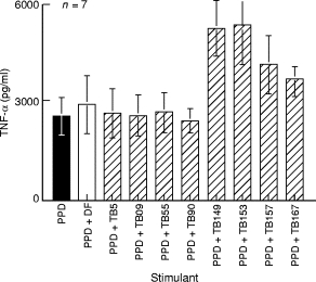
References
-
- Schlesinger LS. Macrophage phagocytosis of virulent but not attenuated strains of Mycobacterium tuberculosis is mediated by mannose receptors in addition to complement receptors. J Immunol. 1993;150:2920–30. - PubMed
-
- Schlesinger LS, Bellinger-Kawahara CG, Payne NR, Horwitz MA. Phagocytosis of Mycobacterium tuberculosis is mediated by human monocyte complement receptor C3. J Immunol. 1990;144:2771–80. - PubMed
-
- Huber HS, Douglas D, Nusbacher J, Kochwa S, Rosenfield RE. IgG subclass specificity of human monocyte receptor sites. Nature. 1971;229:419–20. - PubMed
Publication types
MeSH terms
Substances
Grants and funding
LinkOut - more resources
Full Text Sources
Medical
