Comparative Study
. 2000 Mar;119(3):544-52.
doi: 10.1046/j.1365-2249.2000.01171.x.

In the rheumatoid pannus, anti-filaggrin autoantibodies are produced by local plasma cells and constitute a higher proportion of IgG than in synovial fluid and serum

Affiliations
Comparative Study

In the rheumatoid pannus, anti-filaggrin autoantibodies are produced by local plasma cells and constitute a higher proportion of IgG than in synovial fluid and serum

C Masson-Bessière et al. Clin Exp Immunol. 2000 Mar.

Abstract

IgG anti-filaggrin autoantibodies (AFA) are the most specific serological markers of rheumatoid arthritis (RA). They include the so-called 'anti-keratin antibodies' (AKA) and anti-perinuclear factor (APF), and recognize human epidermal filaggrin and other (pro)filaggrin-related proteins of various epithelial tissues. In this study we demonstrate that AFA are produced in rheumatoid synovial joints. In 31 RA patients, AFA levels were assayed at equal IgG concentrations in paired synovial fluids (SF) and sera. AFA titre-like values determined by indirect immunofluorescence and immunoblotting and AFA concentrations determined by ELISA were non-significantly different in serum and SF, clearly indicating that AFA are not concentrated in SF. In contrast, we demonstrated that AFA are enriched in RA synovial membranes, since the ELISA-determined AFA in low ionic-strength extracts of synovial tissue from four RA patients represented a 7.5-fold higher proportion of total IgG than in paired sera. When small synovial tissue explants from RA patients were cultured for a period of 5 weeks, the profile of IgG and AFA released in the culture supernatants was first consistent with passive diffusion of the tissue-infiltrating IgG (including AFA) over the first day of culture, then with a de novo synthesis of IgG and AFA. Therefore, AFA-secreting plasma cells are present in the synovial tissue of RA patients and AFA can represent a significant proportion of the IgG secreted within the rheumatoid pannus.


Figures

Fig. 1
Fig. 1
Synovial fluid (SF) and serum levels of IgG and anti-filaggrin autoantibodies (AFA) in 31 rheumatoid arthritis (RA) patients. IgG (a) and AFA (b) levels were determined in paired SF and sera obtained from 31 RA patients. IgG concentrations were measured by single radial immunodiffusion. AFA titre-like values were determined by indirect immunofluorescence (IIF) on rat oesophagus cryosections and by immunoblotting on neutral/acidic isoform of human filaggrin. Serum and SF were assayed at the same IgG concentration. AFA concentrations were measured by ELISA using purified neutral/acidic isoform of human filaggrin as coated antigen. Mean values are represented as horizontal bars. The mean IgG concentration was significantly higher in the sera than in the SF (P < 0·00001). The means of AFA in serum and SF were not significantly different whatever the method of assay.
Fig. 2
Fig. 2
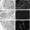
Detection of plasma cells and infiltrating IgG in synovial tissue from rheumatoid arthritis (RA) patients. Paraffin sections of synovial tissue before (A,C) and after (B) 24 h of culture were observed after staining with haematoxylin and eosin. Plasma cells are identified by their eccentric nuclei, large amounts of cytoplasm and perinuclear halo (thin arrows). In (A) plasma cells surround lymphoid follicles (thick arrows); in (B) they are close to blood vessels (arrowheads); in (C) they are interspersed amongst synovial lining cells. Cryosections of synovial tissue before (D) and after (E,F) 24 h of culture were analysed by immunofluorescence using FITC-labelled Fab fragments to human IgG. Before culture the synovial tissue fragments were heavily infiltrated with IgG (D). After 24 h of culture, the level of IgG infiltration was dramatically decreased (E) and clusters of cells exhibiting the nuclear/cytoplasmic ratio typical of plasma cells became distinguishable (F). Bars = 50 μ m.
Fig. 3
Fig. 3
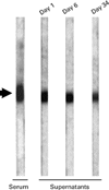
Immunoblotting detection of anti-filaggrin autoantibodies (AFA) produced in vitro by synovial tissue explants. Sixteen 2-mm3 synovial tissue explants from a rheumatoid arthritis (RA) patient (number 3) were cultured separately for a period of 34 days. An immunoblotting reactivity to the neutral/acidic isoform of human filaggrin (arrowhead) is observed in the serum of the patient and in culture supernatants of the synovial explants at days 1, 6 and 34.

Similar articles

Cited by

References

    1. Smiley JD, Sachs C, Ziff M. In vitro synthesis of immunoglobulin by rheumatoid synovial membrane. J Clin Invest. 1968;47:624–32. - PMC - PubMed
    1. Sliwinski AJ, Zvaifler NJ. In vivo synthesis of IgG by rheumatoid synovium. J Lab Clin Med. 1970;76:304–10. - PubMed
    1. Loewi G, Darling J, Howard A. Mononuclear cells from inflammatory joint effusions: electron microscopic appearances and immunoglobulin synthesis. J Rheumatol. 1974;1:34–44. - PubMed
    1. Cush JJ, Lipsky PE. Cellular basis for rheumatoid inflammation. Clin Orthop. 1991;265:9–22. - PubMed
    1. Munthe E, Natvig JB. Immunoglobulin classes, subclasses and complexes of IgG rheumatoid factor in rheumatoid plasma cells. Clin Exp Immunol. 1972;12:55–70. - PMC - PubMed

Publication types