. 2000 Mar;182(6):1499-506.
doi: 10.1128/JB.182.6.1499-1506.2000.

A novel spore peptidoglycan hydrolase of Bacillus cereus: biochemical characterization and nucleotide sequence of the corresponding gene, sleL

Affiliations

A novel spore peptidoglycan hydrolase of Bacillus cereus: biochemical characterization and nucleotide sequence of the corresponding gene, sleL

Y Chen et al. J Bacteriol. 2000 Mar.

Abstract

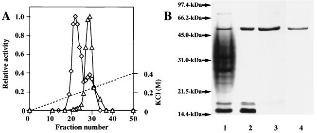
The exudate of germinated spores of B. cereus IFO 13597 in 0.15 M KCl-50 mM potassium phosphate (pH 7.0) contained a spore-lytic enzyme which has substrate specificity for fragmented spore cortex from wild-type organisms (cortical-fragment-lytic enzyme [CFLE]), in addition to a previously characterized germination-specific hydrolase which acts on intact spore cortex (spore cortex-lytic enzyme [SCLE]) (R. Moriyama, S. Kudoh, S. Miyata, S. Nonobe, A. Hattori, and S. Makino, J. Bacteriol. 178:5330-5332, 1996). CFLE was not capable of degrading isolated cortical fragments from spores of Bacillus subtilis ADD1, which lacks muramic acid delta-lactam. This suggests that CFLE cooperates with SCLE in cortex hydrolysis during germination. CFLE was purified in an active form and identified as a 48-kDa protein which functions as an N-acetylglucosaminidase. Immunochemical studies suggested that the mature enzyme is localized on a rather peripheral region of the dormant spore, probably the exterior of the cortex layer. A gene encoding the enzyme, sleL, was cloned in Escherichia coli, and the nucleotide sequence was determined. The gene encodes a protein of 430 amino acids with a deduced molecular weight of 48,136. The N-terminal region contains a repeated motif common to several peptidoglycan binding proteins. Inspection of the data banks showed no similarity of CFLE with N-acetylglucosaminidases found so far, suggesting that CFLE is a novel type of N-acetylglucosaminidase. The B. subtilis genome sequence contains genes, yaaH and ydhD, which encode putative proteins showing similarity to SleL.


Figures

FIG. 1
FIG. 1
Chromatography of CFLE on an SP-Sephadex C25 column and SDS-gel electrophoretic profiles showing purification of the enzyme. (A) Germination exudate (100 ml from 5 g of packed spores) was dialyzed against 60 mM potassium phosphate (pH 8.0) containing 1 mM sodium thioglycollate and put on an SP-Sephadex C25 column. Proteins were eluted with a linear gradient of KCl. Fractions (4 ml each) were collected, and the cortex-lytic activity of each fraction was measured by using cortical fragments (◊) and decoated spores (▵). The broken line shows the molarity of KCl. (B) The CFLE was purified as described in Table 1 and analyzed by 0.1% SDS–12.5% polyacrylamide gel electrophoresis. Approximately 3 to 30 μg of protein was electrophoresed and Coomassie blue stained. Standard proteins were run with samples in each gel, and the time of electrophoresis for lane 3 was not the same as those for other lanes. Lanes: 1, germination exudate; 2, fractions containing CFLE activity eluted from an SP-Sephadex C25 column; 3, fractions containing CFLE activity eluted from a hydroxyapatite column; 4, Superose 12 column-purified enzyme. The standard proteins run were rabbit muscle phosphorylase b (97.4 kDa), bovine serum albumin (66.2 kDa), ovalbumin (45.0 kDa), bovine carbonic anhydrase (31.0 kDa), soybean trypsin inhibitor (21.5 kDa), and egg white lysozyme (14.4 kDa).
FIG. 2
FIG. 2
Release of reducing groups during digestion of cortical fragments with CFLE. Cortical fragments of C. perfringens spores (8.0 mg) were suspended in 2.5 ml of 0.1 M potassium phosphate (pH 6.0). CFLE (3.0 U; 25 μl) was added to the suspension, and the digestion was performed at 32°C. Aliquots were taken at the indicated times for determination of the turbidity at 600 nm in a cell of 1 mm light path (□), the release of reducing sugars (○), and the release of amino groups (▵). No decrease in turbidity was seen when heat-denatured enzyme was used (■). The same results were obtained when B. subtilis spore cortical fragments were used as a substrate.
FIG. 3
FIG. 3
Effects of pH, temperature, and NaCl concentration on the activity of CFLE. The CFLE activity is shown relative to the maximum activity. (A) Enzyme (5 μl with 0.08 U) in 5 mM Tris-HCl (pH 7.6) was mixed with 135 μl of 0.1 M buffer solutions to obtain the indicated pH. After incubation at 32°C for 10 min, 5 μl of C. perfringens cortical fragments was added and the enzyme activity was measured. The following buffers were used: CH3COOH-CH3COONa (□), Na2HPO4-NaH2PO4 (◊), and NaHCO3-Na2CO3 (○). (B) Enzyme (5 μl with 0.08 U) in 5 mM Tris-HCl (pH 7.6) was mixed with 135 μl of 5 mM Tris-HCl (pH 7.6) containing 0.1 M NaCl and incubated for 30 min at the indicated temperature. Then 5 μl of cortical fragments was added, and the residual activity was assayed at 32°C. (C) Enzyme (5 μl with 0.08 U) in 5 mM Tris-HCl (pH 7.6) was mixed with 135 μl of 5 mM Tris-HCl (pH 7.6) containing the desired concentration of NaCl. After incubation at 32°C for 10 min, 5 μl of cortical fragments was added and the enzyme activity was measured. Similar results were obtained when KCl was replaced with NaCl.
FIG. 4
FIG. 4
Immunological detection of CFLE-related proteins in dormant spores and vegetative cells. Spores and vegetative cells were disrupted in and extracted with 0.15 M KCl–50 mM potassium phosphate (pH 7.0), and the coat fraction was recovered as described in the text. The germination exudate, the spore extracts, and the coat fraction were subjected to SDS-polyacrylamide gel electrophoresis followed by immunoblotting. Before electrophoresis, all the samples were dialyzed against 10 mM Tris-HCl (pH 8.0) containing 0.2% SDS and 5 mM 2-mercaptoethanol, and approximately 3 to 30 μg of protein was loaded on the gel. Lanes: 1, purified CFLE; 2, germination exudate; 3, extract made from disrupted dormant spores; 4, the same extract as in lane 3 but pretreated with anti-CFLE serum; 5, extract made from disrupted vegetative cells; 6, coat fraction; 7, extract made from disrupted decoated spores. Prior to electrophoresis, the CFLE activity of the samples was examined. Symbols: +, positive hydrolytic activity; −, no hydrolytic activity.
FIG. 5
FIG. 5
Nucleotide sequence of B. cereus chromosomal DNA between two oligonucleotide primers, SleLF4 and SleLR2, and deduced amino acid sequences. The deduced amino acid sequences of orf1 (nucleotides 1 to 210), sleL (nucleotides 371 to 1660), and orf3 (nucleotides 1695 to 1860) are given below the nucleotide sequence. The numbers of the nucleotides and amino acids are shown on the right. A candidate for a putative ribosome-binding site (RBS) is indicated by shading. The asterisk indicates a termination codon. The bold-underlined amino acid sequence shows the N-terminal sequence of CFLE (20 residues), and the thin-underlined sequences show peptides A, B, C, D, and E derived from the purified enzyme by digestion with trypsin. An inverted repeat is indicated by arrows.
FIG. 6
FIG. 6
Comparison of the amino acid sequence of B. cereus CFLE (SleL) with those of YaaH and YdhD of B. subtilis. The amino acid sequences are numbered on the left. Identical amino acids in two or three proteins are shown by a black background. Dashes are introduced to obtain a better alignment of the sequences.

References

    1. Amutha B, Khire J M, Khan M S. Active site characterization of the exo-N-acetyl-β-d-glucosaminidase from thermotolerant Bacillus sp. NCIM 5120: involvement of tryptophan, histidine and carboxylate residues in catalytic activity. Biochim Biophys Acta. 1999;1427:121–132. - PubMed
    1. Atrih A, Bacher G, Körner R, Allmaier G, Foster S J. Structural analysis of Bacillus megaterium KM spore peptidoglycan and its dynamics during germination. Microbiology. 1999;145:1033–1041. - PubMed
    1. Atrih A, Zöllner P, Allmaier G, Williamson M P, Foster S J. Structural analysis of Bacillus subtilis 168 endospore peptidoglycan and its role during differentiation. J Bacteriol. 1996;178:6173–6183. - PMC - PubMed
    1. Atrih A, Zöllner P, Allmaier G, Williamson M P, Foster S J. Peptidoglycan structural dynamics during germination of Bacillus subtilis 168 endospores. J Bacteriol. 1998;180:4603–4612. - PMC - PubMed
    1. Bagyan I, Setlow B, Setlow P. New small, acid-soluble proteins unique to spores of Bacillus subtilis: identification of the coding genes and regulation and function of two of these genes. J Bacteriol. 1998;180:6704–6712. - PMC - PubMed

Publication types

MeSH terms

Associated data

LinkOut - more resources