. 2000 Mar;182(6):1659-70.
doi: 10.1128/JB.182.6.1659-1670.2000.

Analysis of Escherichia coli RecA interactions with LexA, lambda CI, and UmuD by site-directed mutagenesis of recA

Affiliations

Analysis of Escherichia coli RecA interactions with LexA, lambda CI, and UmuD by site-directed mutagenesis of recA

J A Mustard et al. J Bacteriol. 2000 Mar.

Abstract

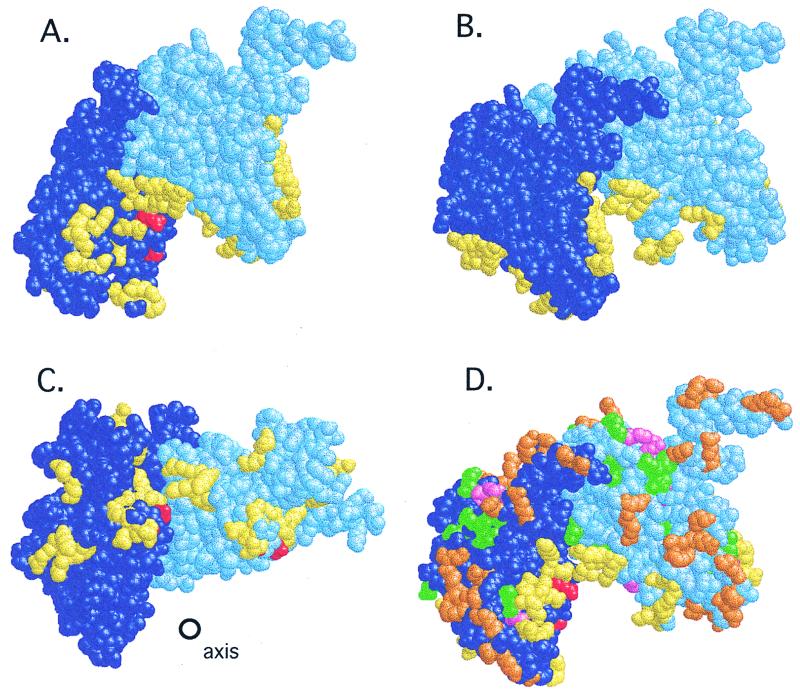
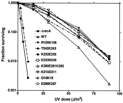
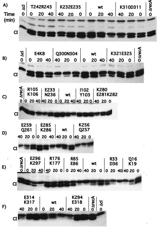
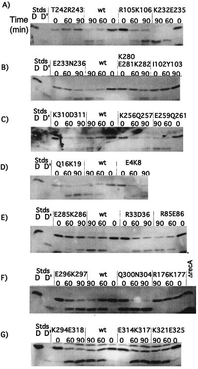
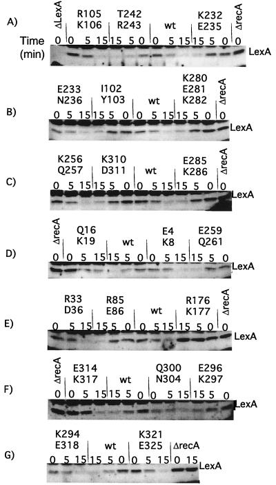
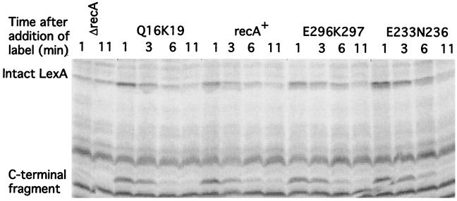
An early event in the induction of the SOS system of Escherichia coli is RecA-mediated cleavage of the LexA repressor. RecA acts indirectly as a coprotease to stimulate repressor self-cleavage, presumably by forming a complex with LexA. How complex formation leads to cleavage is not known. As an approach to this question, it would be desirable to identify the protein-protein interaction sites on each protein. It was previously proposed that LexA and other cleavable substrates, such as phage lambda CI repressor and E. coli UmuD, bind to a cleft located between two RecA monomers in the crystal structure. To test this model, and to map the interface between RecA and its substrates, we carried out alanine-scanning mutagenesis of RecA. Twenty double mutations were made, and cells carrying them were characterized for RecA-dependent repair functions and for coprotease activity towards LexA, lambda CI, and UmuD. One mutation in the cleft region had partial defects in cleavage of CI and (as expected from previous data) of UmuD. Two mutations in the cleft region conferred constitutive cleavage towards CI but not towards LexA or UmuD. By contrast, no mutations in the cleft region or elsewhere in RecA were found to specifically impair the cleavage of LexA. Our data are consistent with binding of CI and UmuD to the cleft between two RecA monomers but do not provide support for the model in which LexA binds in this cleft.


Figures

FIG. 1
FIG. 1
Location of site-directed changes in the RecA structure. Shown are two adjacent monomers from the RecA filament (see the text), colored dark and light blue. In the view in panel D, the helix axis lies directly behind the interface between the two monomers and is oriented vertically (not shown). The views in panels A and B are that of panel D rotated by 30° or −30° along the y axis; that in panel C is the view of panel D rotated −90° along the y axis so that the cleft is viewed from beneath; the helix axis is shown. (A to C) Mutational changes in the cleft. Residues Arg243 and Gly229 are shown in red; residues changed in the cleft (identified in Table 3) are shown in yellow. (D) Locations of other mutations and changes on the outer surface of the filament. Mutations destroying RecA function (listed in Table 2) are shown in pink; those that leave LexA cleavage intact (Table 2) are shown in green; changes made in the present work (Table 3) are shown in orange; yellow and red are as in panels A to C. RasMol scripts generating these views are available at http://www.biochem.arizona.edu/Little/Littlelab.htm.
FIG. 2
FIG. 2
UV survival curves of wild type and selected mutants. Derivatives of JL2460 carrying plasmids with the indicated recA alleles are shown; the ΔrecA host carried pBR322. Cells were grown and irradiated with various doses of UV light as described in Materials and Methods; the fraction surviving was determined as described. The legend inset in the graph identifies the recA allele carried by each strain.
FIG. 3
FIG. 3
In vivo cleavage of λ CI repressor by mutant RecA proteins. Derivatives of JAM267 carrying pTrecA220 or mutant derivatives were treated as described in Materials and Methods. The strain marked ΔrecA carried pBR322. Each panel represents the results of a representative experiment. For each mutant strain, a time course is shown. The lanes marked “0” contain samples taken without treatment; those marked “20” and “40” were taken 20 and 40 min after the addition of nalidixic acid (50 μg/ml). The samples were analyzed by Western blotting with antibody against λ CI (see Materials and Methods). The name of the mutation is shown above each set of samples. The location of intact CI is shown; the band just above CI is a cross-reacting protein. wt, wild type.
FIG. 4
FIG. 4
In vivo cleavage of UmuD by mutant RecA proteins. Derivatives of JL2460 carrying pTrecA220 or mutant derivatives were grown and treated as described in Materials and Methods; the strain marked ΔrecA carried pBR322. Each panel represents the results of a representative experiment. For each mutant strain, a time course is shown. Lanes marked “D” and “D′” are standards (Stds) from cells overproducing UmuD and UmuD′, respectively, and serve as markers. The lanes marked “0” contain samples taken without treatment; those marked “60” and “90” were taken 60 and 90 min after addition of nalidixic acid (50 μg/ml). The samples were analyzed by Western blotting with antibody to UmuD, as described in Materials and Methods. The name of the mutation is shown above each set of samples. wt, wild type.
FIG. 5
FIG. 5
In vivo cleavage of LexA by mutant RecA proteins. Derivatives of strain JAM444 carrying pTrecA220 or mutant derivatives were grown and treated as described in Materials and Methods; the strain marked ΔrecA carried pBR322. Each panel represents the results of a representative experiment. For each mutant strain, a time course is shown. The lanes marked “0” contain samples taken without treatment; those marked “5” and “15” were taken 5 and 15 min after the addition of nalidixic acid (50 μg/ml). Samples were analyzed by Western blotting with antibody to LexA, as described in Materials and Methods. The name of the mutation is shown above each set of samples. The location of intact LexA is shown; the other bands are proteins that cross-reacted with the LexA antibody. Cleavage fragments are not shown; their levels are uninformative, since LexA cleavage fragments are unstable (27) (for an example, cf. Fig. 6). As described in the text, several mutants had less basal cleavage than the wild type. The amount of basal LexA cleavage did not appear to depend on the amount of RecA mutant protein. Q16K19 had very low levels of basal cleavage yet had higher levels of mutant RecA. On the other hand, K294E318 protein was present at very low levels and yet had the same amount of basal cleavage of LexA as the wild type (wt). The fact that the amount of basal cleavage did not correlate with the amount of RecA suggests that the extent of basal cleavage was limited by the amount of signal in the cell and/or by the ability of RecA protein to become activated by it.
FIG. 6
FIG. 6
Pulse-chase analysis of LexA cleavage for selected mutants. Derivatives of JL783 carrying pTrecA220 or mutant derivatives were grown as described in Materials and Methods; the strain marked ΔrecA carried pBR322. The cultures were treated with nalidixic acid for 30 min, followed by the addition of [35S]methionine (50 μCi/ml) and sampling at the times indicated. The identity of each set of samples is indicated above the gel. The samples were precipitated with antibody to LexA and analyzed as described in the text. The locations of intact LexA and the C-terminal cleavage product are indicated. The other bands are cross-reacting species also precipitated by the antibody. In the ΔrecA strain, less LexA is made, because LexA negatively autoregulates its own expression.

Similar articles

Cited by

References

    1. Alexseyev A A, Bakhlanova I V, Zaitsev E N, Lanzov V A. Genetic characteristics of new recA mutants of Escherichia coli K-12. J Bacteriol. 1996;178:2018–2024. - PMC - PubMed
    1. Backman K, Ptashne M. Maximizing gene expression on a plasmid using recombination in vitro. Cell. 1978;13:65–71. - PubMed
    1. Bailone A, Levine A, Devoret R. Inactivation of prophage λ repressor in vivo. J Mol Biol. 1979;131:553–572. - PubMed
    1. Bolivar F, Rodriguez R L, Greene P J, Betlach M C, Heyneker H L, Boyer H W, Crosa J H, Falkow S. Construction and characterization of new cloning vehicles. II. A multipurpose cloning system. Gene. 1977;2:95–113. - PubMed
    1. Churchward G, Belin D, Nagamine Y. A pSC101-derived plasmid which shows no sequence homology to other commonly used cloning vectors. Gene. 1984;31:165–171. - PubMed

Publication types

MeSH terms

LinkOut - more resources