Role of nitric oxide- and vasoactive intestinal polypeptide-containing neurones in human gastric fundus strip relaxations
- PMID: 10694197
- PMCID: PMC1621112
- DOI: 10.1038/sj.bjp.0702977
Role of nitric oxide- and vasoactive intestinal polypeptide-containing neurones in human gastric fundus strip relaxations
Abstract
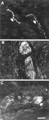
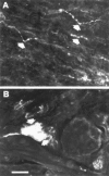
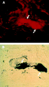
The morphological pattern and motor correlates of nitric oxide (NO) and vasoactive intestinal polypeptide (VIP) innervation in the human isolated gastric fundus was explored. By using the nicotinamide adenine dinucleotide phosphate hydrogen (NADPH)-diaphorase and specific rabbit polyclonal NO-synthase (NOS) and VIP antisera, NOS- and VIP-containing varicose nerve fibres were identified throughout the muscle layer or wrapping ganglion cell bodies of the myenteric plexus. NOS-immunoreactive (IR) neural cell bodies were more abundant than those positive for VIP-IR. The majority of myenteric neurones containing VIP coexpressed NADPH-diaphorase. Electrical stimulation of fundus strips caused frequency-dependent NANC relaxations. N(G)-nitro-L-arginine (L-NOARG: 300 microM) enhanced the basal tone, abolished relaxations to 0.3 - 3 Hz (5 s) and those to 1 Hz (5 min), markedly reduced ( approximately 50%) those elicited by 10 - 50 Hz, and unmasked or potentiated excitatory cholinergic responses at frequencies > or =1 Hz. L-NOARG-resistant relaxations were virtually abolished by VIP (100 nM) desensitization at all frequencies. Relaxations to graded low mechanical distension (< or =1 g) were insensitive to tetrodotoxin (TTX: 1 microM) and L-NOARG (300 microM), while those to higher distensions (2 g) were slightly inhibited by both agents to the same extent ( approximately 25%). In the human gastric fundus, NOS- and VIP immunoreactivities are colocalized in the majority of myenteric neurones. NO and VIP mediate electrically evoked relaxations: low frequency stimulation, irrespective of the duration, caused NO release only, whereas shortlasting stimulation at high frequencies induced NO and VIP release. Relaxations to graded mechanical distension were mostly due to passive viscoelastic properties, with a slight NO-mediated neurogenic component at 2 g distension. The difference between NO and VIP release suggests that in human fundus accommodation is initiated by NO. British Journal of Pharmacology (2000) 129, 12 - 20
Figures


Similar articles
-
Study of NO and VIP as non-adrenergic non-cholinergic neurotransmitters in the pig gastric fundus.Br J Pharmacol. 1995 Oct;116(3):2017-26. doi: 10.1111/j.1476-5381.1995.tb16406.x. Br J Pharmacol. 1995. PMID: 8640340 Free PMC article.
-
Involvement of nitric oxide in non-adrenergic non-cholinergic relaxation and action of vasoactive intestinal polypeptide in circular muscle strips of the rat gastric fundus.Pharmacol Res. 2001 Sep;44(3):221-8. doi: 10.1006/phrs.2001.0844. Pharmacol Res. 2001. PMID: 11529689
-
Nitric oxide, and not vasoactive intestinal peptide, as the main neurotransmitter of vagally induced relaxation of the guinea pig stomach.Br J Pharmacol. 1994 Dec;113(4):1197-202. doi: 10.1111/j.1476-5381.1994.tb17124.x. Br J Pharmacol. 1994. PMID: 7534182 Free PMC article.
-
Interaction of NO and VIP in gastrointestinal smooth muscle relaxation.Curr Pharm Des. 2004;10(20):2483-97. doi: 10.2174/1381612043383890. Curr Pharm Des. 2004. PMID: 15320758 Review.
-
Non-adrenergic non-cholinergic relaxation of the rat stomach.Gen Pharmacol. 1998 Nov;31(5):697-703. doi: 10.1016/s0306-3623(98)00096-2. Gen Pharmacol. 1998. PMID: 9809465 Review.
Cited by
-
NANC inhibitory neurotransmission in mouse isolated stomach: involvement of nitric oxide, ATP and vasoactive intestinal polypeptide.Br J Pharmacol. 2003 Sep;140(2):431-7. doi: 10.1038/sj.bjp.0705431. Epub 2003 Aug 11. Br J Pharmacol. 2003. PMID: 12970100 Free PMC article.
-
Accelerated Electron Ionization-Induced Changes in the Myenteric Plexus of the Rat Stomach.Int J Mol Sci. 2024 Jun 20;25(12):6807. doi: 10.3390/ijms25126807. Int J Mol Sci. 2024. PMID: 38928511 Free PMC article.
-
Serum from achalasia patients alters neurochemical coding in the myenteric plexus and nitric oxide mediated motor response in normal human fundus.Gut. 2006 Mar;55(3):319-26. doi: 10.1136/gut.2005.070011. Epub 2005 Aug 16. Gut. 2006. PMID: 16105888 Free PMC article.
-
Relaxation patterns of human gastric corporal smooth muscle by cyclic nucleotides producing agents.Korean J Physiol Pharmacol. 2009 Dec;13(6):503-10. doi: 10.4196/kjpp.2009.13.6.503. Epub 2009 Dec 31. Korean J Physiol Pharmacol. 2009. PMID: 20054499 Free PMC article.
-
Heme deficiency of soluble guanylate cyclase induces gastroparesis.Neurogastroenterol Motil. 2013 May;25(5):e339-52. doi: 10.1111/nmo.12120. Epub 2013 Mar 28. Neurogastroenterol Motil. 2013. PMID: 23551931 Free PMC article.
References
-
- ABRAHAMSSON H. Studies on the inhibitory nervous control of gastric motility. Acta Physiol. Scand. 1973;390:1–38. - PubMed
-
- ALLESCHER H.D., TOUGAS G., VERGARA P., LU S., DANIEL E.E. Nitric oxide as a putative nonadrenergic noncholinergic inhibitory transmitter in the canine pylorus in vivo. Am. J. Physiol. 1992;262:G695–G702. - PubMed
-
- BACCARI M.C., BERTINI M., CALAMAI F. Effects of L-NG-nitro arginine on cholinergic transmission in the gastric muscle of the rabbit. Neuroreport. 1993;4:1102–1104. - PubMed
-
- BARBIER A.J., LEFEBVRE R.A. Involvement of the L-arginine: nitric oxide pathway in nonadrenergic noncholinergic relaxation of the cat gastric fundus. J. Pharmacol. Exp. Ther. 1993;266:172–178. - PubMed
-
- BARTFAI T., IVERFELDT K., FISONE G. Regulation of the release of coexisting neurotransmitters. Ann. Rev. Pharmacol. Toxicol. 1988;28:285–310. - PubMed
Publication types
MeSH terms
Substances
Grants and funding
LinkOut - more resources
Full Text Sources

