Cervical diskography: analysis of provoked responses at C2-C3, C3-C4, and C4-C5
- PMID: 10696007
- PMCID: PMC7975353
Cervical diskography: analysis of provoked responses at C2-C3, C3-C4, and C4-C5
Abstract
Background and purpose: Previous authors have described the locations of provoked responses to cervical diskography from C3-C4 to C6-C7, but we have found no description of the findings at C2-C3. This study was undertaken to analyze the sensations provoked during cervical diskography at C2-C3 and to compare the results with those provoked at C3-C4 and C4-C5.
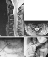
Methods: The locations of diskographically provoked responses from 40 consecutive patients who had undergone C2-C3, C3-C4, and C4-C5 diskography were analyzed. Only intensely painful (> or = 7/10) and concordant responses were considered. Disk morphology on MR images and diskograms was also compared with the provoked responses.
Results: Eighteen subjects described either unilateral (n = 10) or bilateral (usually asymmetric) (n = 8) concordant pain at the craniovertebral junction in response to C2-C3 diskography. Nine subjects described either unilateral (n = 5) or bilateral (n = 4) neck pain during injection. Cephalalgia or head pain was provoked in 19 subjects, seven bilaterally. Four subjects described either unilateral (n = 3) or bilateral (n = 1) trapezius muscle and/or shoulder pain. Preliminary MR studies were not helpful, as most C2-C3 disks either appeared normal or exhibited nonspecific signs of degeneration. All disks exhibited either fissuring or extradiskal leakage of contrast material at diskography, regardless of the response provoked.
Conclusion: Diskography at C2-C3 and C3-C4 frequently produces pain sensations in the head, craniovertebral junction, and neck. There is no correlation between C2-C3 disk morphology and the diskographically provoked response.
Figures


Comment in
-
Diskography: science and the ad hoc hypothesis.AJNR Am J Neuroradiol. 2000 Feb;21(2):241-2. AJNR Am J Neuroradiol. 2000. PMID: 10696000 Free PMC article. No abstract available.
References
-
- Barnsley L, Lord S, Bogduk N. Clinical review: whiplash injury. Pain 1994;58:283-307 - PubMed
-
- Bogduk N. Innervation and pain patterns in the cervical spine. Clin Phys Ther 1988;17:1-13
-
- Bogduk N. The anatomy and pathophysiology of whiplash. Clin Biomech 1986;1:92-101 - PubMed
-
- Bogduk N, Windsor M, Inglis A. The innervation of the cervical intervertebral discs. Spine 1988;13:2-8 - PubMed
MeSH terms
LinkOut - more resources
Full Text Sources
Medical
Miscellaneous
