

Registration of three-dimensional MR and CT studies of the cervical spine
Affiliations
- PMID: 10696009
- PMCID: PMC7975327
Item in Clipboard
Registration of three-dimensional MR and CT studies of the cervical spine
AJNR Am J Neuroradiol.
2000 Feb.
Abstract
A three-dimensional image registration technique for CT and MR studies of the cervical spine was evaluated for feasibility and efficacy. Registration by means of external fiducial markers was slightly more accurate than registration by anatomic landmarks. The interrelationships between bony (eg, neural foramina) and soft tissue structures (eg, nerve roots) in the cervical spine were more conspicuous on registered images than on conventional displays. Registration of CT and MR images may be used to examine more precisely the relationships between bony and soft tissue structures of the cervical spine.
Figures
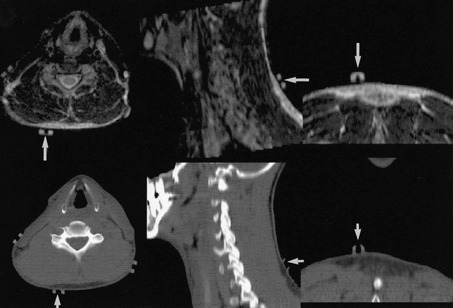
Location of external fiducials on orthogonal CT scans and MR images at a comparable level of the cervical spine. The external fiducial landmark (arrows) is first located on the axial view of the individual imaging studies and then compared with the location on the reformatted sagittal and coronal sections through the image volume
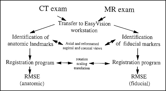
Diagram of the registration process
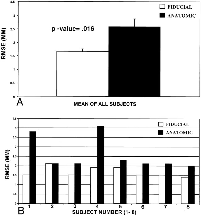
A, Bar graph shows that the RMSE value of the fiducial registration in all eight subjects is less than that of the anatomic registration (error bars represent SEM). B, Comparative profile of anatomic and fiducial RMSE values for each subject. Subject 2 has identical RMSE values for both the anatomic and fiducial registrations.

Multiple axial images from one subject (subject 2 from fig 3B) include corresponding MR image (left), CT scan (middle), and registered image (right) of the cervical spine at the level of the neural foramina (two levels). This was the format used by the three neuroradiologists to compare the clarity of conventional images (MR and CT studies side by side) with that of the registered images. The relationships between the margins of the neural foramina (solid arrow) and spinal canal (open arrow) and their contents, respectively, are more conspicuously delineated on the fused image. The images were registered using external fiducial landmarks
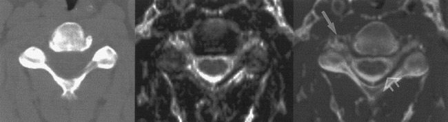
Magnified view of the axial images of the cervical spine at a comparable level (CT scan, left; MR image, center; registered image, right). The relationship between the margins of the neural foramina (solid arrow) and spinal canal (open arrow) and their contents is more conspicuously delineated on the registered image. The images were registered using external fiducial landmarks. Subject 5 from figure 3B is represented
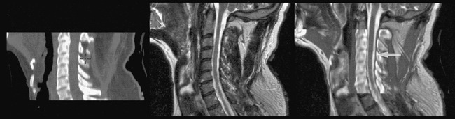
Corresponding midsagittal CT (left), MR (middle), and registered (right) images of the cervical spine show proper alignment and the relationship between the margin of the spinal canal (solid arrow) and its contents. The images were registered using external fiducial landmarks. Subject 4 from figure 3B is represented
References
-
- Hill DLG, Hawkes DJ, Gleeson MJ, et al. Accurate frameless registration of MR and CT images of the head: applications in surgery and radiotherapy planning. Radiology 1994;191:447-454 - PubMed
-
- Hill DLG, Hawkes DJ, Crossmann JE, et al. Registration of MR and CT images for skull base surgery using point-like anatomical features. Br J Radiol 1991;64:1030-1035 - PubMed
-
- Hill DLG, Hawkes DJ, Hussain Z, et al. Accurate combination of CT and MR data of the head: validation and applications in surgical and therapy planning. Comput Med Imaging 1993;17:357-362 - PubMed
-
- Gandhe AJ, Hill DLG, Studholm C, et al. Combined and three-dimensional rendered multimodal data for planning cranial base surgery: a prospective evaluation. Neurosurgery 1994;35:463-471 - PubMed
-
- Arun KS, Huang TS, Blostein SD. Least square fitting of two 3D point sets. IEEE Trans Pattern Anal Mach Intell 1987;9:698-700 - PubMed
MeSH terms
LinkOut - more resources
Full Text Sources
Medical