

Second branchial cleft cysts: variability of sonographic appearances in adult cases
- PMID: 10696015
- PMCID: PMC7975337
Second branchial cleft cysts: variability of sonographic appearances in adult cases
Abstract
Background and purpose: Previous reports have suggested that second branchial cleft cysts (BCCs) appear on sonograms as well-defined, cystic masses with thin walls and posterior enhancement. Previous CT and MR imaging findings, however, have indicated heterogeneity of these masses, and, in our experience, sonography also shows a similar variable appearance. In this communication, we report the cases of 17 patients with second BCCs and document the variability of sonographic patterns.
Methods: The sonograms of 17 adults with second BCCs were reviewed. Only patients with surgical or cytologic evidence of BCCs were included in this study. The features evaluated were the location, internal echogenicity, posterior enhancement, and presence of septa and fistulous tract.
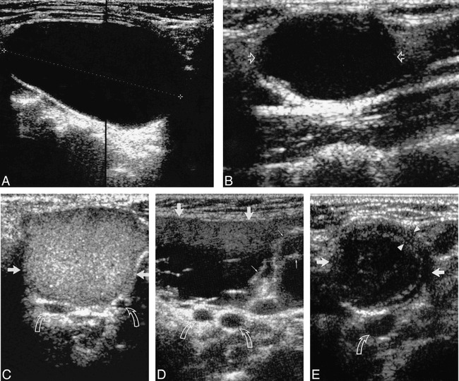
Results: Four patterns of second BCCs were identified: anechoic (41%), homogeneously hypoechoic with internal debris (23.5%), pseudosolid (12%), and heterogeneous (23.5%). The majority (70%) showed posterior enhancement. All were situated in their classical location, posterior to the submandibular gland, superficial to the carotid artery and internal jugular vein, and closely related to the medial and anterior margin of the sternomastoid muscle. Fourteen (82%) of the 17 BCCs had imperceptible walls, and all were well defined. For none of the patients was a fistulous tract revealed by sonography; the presence of internal septations was revealed for three patients.
Conclusion: As previously suggested by CT and MR imaging findings, sonography reinforces that second BCCs in adults are not simple cysts but have a complex sonographic pattern ranging from a typical anechoic to a pseudosolid appearance.
Figures
References
-
- Hyndman OR, Light G. The branchial apparatus. Arch Surg 1929;19:410-452
-
- Shedden WM. Branchial cysts and fistulae. N Engl J Med 1931;205:800-811
-
- Neel HB, Pemberton J. Lateral cervical cysts and fistulas. Surgery 1945;18:267-286
-
- Weismann JL. Nonnodal masses of the neck. In: Som PM, Curtin HD, eds. Head & Neck Imaging. 3rd ed. St. Louis: Mosby-Year Book; 1996:794-822
-
- Harnsberger HR. Cystic masses of the head and neck: rare lesions with characteristic radiologic features. In: Harnsberger RH, ed. Handbook of Head & Neck Imaging. 2nd ed. St. Louis: Mosby Year-Book; 1995:199-223
Publication types
MeSH terms
LinkOut - more resources
Full Text Sources
Research Materials